 Movie
Movie Controller

[English] 日本語
 Yorodumi
- PDB-3d61: Crystal Structure Analysis of 1,5-alpha-arabinanase catalytic mut... -
+ Open data

- Basic information
| Entry | Database: PDB / ID: 3d61 | |||||||||
|---|---|---|---|---|---|---|---|---|---|---|
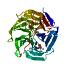
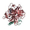
| Title | Crystal Structure Analysis of 1,5-alpha-arabinanase catalytic mutant (AbnBD147A) complexed to arabinobiose | |||||||||
 Components Components | Intracellular arabinanase | |||||||||
 Keywords Keywords |  HYDROLASE / Arabinanase / Glycosyl Hydrolase / beta-propeller / Geobacillus stearothermophilus HYDROLASE / Arabinanase / Glycosyl Hydrolase / beta-propeller / Geobacillus stearothermophilus | |||||||||
| Function / homology |  Function and homology information Function and homology informationarabinan endo-1,5-alpha-L-arabinanase / arabinan endo-1,5-alpha-L-arabinosidase activity / arabinan catabolic process / metal ion binding / cytoplasmSimilarity search - Function | |||||||||
| Biological species |   Geobacillus stearothermophilus (bacteria) Geobacillus stearothermophilus (bacteria) | |||||||||
| Method | X-RAY DIFFRACTION / MOLECULAR REPLACEMENT / Resolution: 1.95 Å | |||||||||
 Authors Authors | Alhassid, A. / Ben David, A. / Shoham, Y. / Shoham, G. | |||||||||
 Citation Citation | Journal: Biochem.J. / Year: 2009 Title: Crystal structure of an inverting GH 43 1,5-alpha-L-arabinanase from Geobacillus stearothermophilus complexed with its substrate Authors: Alhassid, A. / Ben-David, A. / Tabachnikov, O. / Libster, D. / Naveh, E. / Zolotnitsky, G. / Shoham, Y. / Shoham, G. | |||||||||
| History |
|
- Structure visualization
| Structure viewer | Molecule: MolmilJmol/JSmol |
|---|
- Downloads & links
-Download
| PDBx/mmCIF format | 3d61.cif.gz | 82.8 KB | Display | PDBx/mmCIF format |
|---|---|---|---|---|
| PDB format | pdb3d61.ent.gz | 59.8 KB | Display | PDB format |
| PDBx/mmJSON format | 3d61.json.gz | Tree view | PDBx/mmJSON format | |
| Others |  Other downloads Other downloads |
-Validation report
| Arichive directory | https://data.pdbj.org/pub/pdb/validation_reports/d6/3d61ftp://data.pdbj.org/pub/pdb/validation_reports/d6/3d61 | HTTPS FTP |
|---|
-Related structure data
| Related structure data | 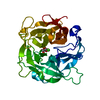 3cu9SC  3d5yC 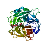 3d5zC 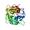 3d60C S: Starting model for refinement C: citing same article ( |
|---|---|
| Similar structure data |


-Links
 PDBj
PDBj- Assembly
| Deposited unit | 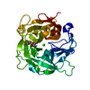
| ||||||||
|---|---|---|---|---|---|---|---|---|---|
| 1 |
| ||||||||
| 2 | 
| ||||||||
| Unit cell |
|
-Components
| #1: Protein | Mass: 35616.605 Da / Num. of mol.: 1 / Mutation: D147A Source method: isolated from a genetically manipulated source Source: (gene. exp.) Geobacillus stearothermophilus (bacteria)Strain: T-6 / Gene: abn / Plasmid: pET9d / Production host: Escherichia coli (E. coli) / Strain (production host): BL21(DE3)References: UniProt: B3EYM8, arabinan endo-1,5-alpha-L-arabinanase |
|---|---|
| #2: Polysaccharide | alpha-L-arabinofuranose-(1-5)-beta-L-arabinofuranose / Mass: 282.245 Da / Num. of mol.: 1 Source method: isolated from a genetically manipulated source |
| #3: Chemical | ChemComp-CA /   Mass: 40.078 Da / Num. of mol.: 1 / Source method: obtained synthetically / Formula: Ca Mass: 40.078 Da / Num. of mol.: 1 / Source method: obtained synthetically / Formula: Ca |
| #4: Water | ChemComp-HOH / Water Mass: 18.015 Da / Num. of mol.: 227 / Source method: isolated from a natural source / Formula: H2O Mass: 18.015 Da / Num. of mol.: 227 / Source method: isolated from a natural source / Formula: H2O |
-Experimental details
-Experiment
| Experiment | Method: X-RAY DIFFRACTION / Number of used crystals: 1 |
|---|
- Sample preparation
| Crystal | Density Matthews: 2.06 Å3/Da / Density % sol: 40.29 % |
|---|---|
| Crystal grow | Temperature: 295 K / Method: vapor diffusion, hanging drop, cocrystaliization / pH: 8 Details: 1.6M lithium sulfate, 0.1M Tris buffer pH 8, 0.5mM arabinobiose, Vapor diffusion, Hanging drop, Cocrystaliization, temperature 295K |
-Data collection
| Diffraction | Mean temperature: 100 K |
|---|---|
| Diffraction source | Source: ROTATING ANODE / Type: RIGAKU RU300 / Wavelength: 1.54 Å |
| Detector | Type: RIGAKU RAXIS IV / Detector: IMAGE PLATE / Date: Jan 7, 2007 |
| Radiation | Monochromator: Ni FILTER / Protocol: SINGLE WAVELENGTH / Monochromatic (M) / Laue (L): M / Scattering type: x-ray |
| Radiation wavelength | Wavelength: 1.54 Å / Relative weight: 1 |
| Reflection | Resolution: 1.95→50 Å / Num. all: 21840 / Num. obs: 21215 / Redundancy: 5.6 % / Biso Wilson estimate: 14 Å2 / Rmerge(I) obs: 0.082 / Rsym value: 0.067 |
- Processing
| Software |
| ||||||||||||||||||||||||||||||||||||||||||||||||||||||||||||||||||||||||||||||||
|---|---|---|---|---|---|---|---|---|---|---|---|---|---|---|---|---|---|---|---|---|---|---|---|---|---|---|---|---|---|---|---|---|---|---|---|---|---|---|---|---|---|---|---|---|---|---|---|---|---|---|---|---|---|---|---|---|---|---|---|---|---|---|---|---|---|---|---|---|---|---|---|---|---|---|---|---|---|---|---|---|---|
| Refinement | Method to determine structure: MOLECULAR REPLACEMENT Starting model: PDB ENTRY 3CU9 Resolution: 1.95→28.99 Å / Rfactor Rfree error: 0.005 / Data cutoff high absF: 377765.09 / Data cutoff low absF: 0 / Isotropic thermal model: RESTRAINED / Cross valid method: THROUGHOUT / σ(F): 0
| ||||||||||||||||||||||||||||||||||||||||||||||||||||||||||||||||||||||||||||||||
| Solvent computation | Solvent model: FLAT MODEL / Bsol: 39.1349 Å2 / ksol: 0.362629 e/Å3 | ||||||||||||||||||||||||||||||||||||||||||||||||||||||||||||||||||||||||||||||||
| Displacement parameters | Biso mean: 22.3 Å2
| ||||||||||||||||||||||||||||||||||||||||||||||||||||||||||||||||||||||||||||||||
| Refine analyze |
| ||||||||||||||||||||||||||||||||||||||||||||||||||||||||||||||||||||||||||||||||
| Refinement step | Cycle: LAST / Resolution: 1.95→28.99 Å
| ||||||||||||||||||||||||||||||||||||||||||||||||||||||||||||||||||||||||||||||||
| Refine LS restraints |
| ||||||||||||||||||||||||||||||||||||||||||||||||||||||||||||||||||||||||||||||||
| LS refinement shell | Resolution: 1.95→2.07 Å / Rfactor Rfree error: 0.015 / Total num. of bins used: 6
| ||||||||||||||||||||||||||||||||||||||||||||||||||||||||||||||||||||||||||||||||
| Xplor file |
|
