 Movie
Movie Controller

[English] 日本語
 Yorodumi
- PDB-3d5y: High resolution crystal structure of 1,5-alpha-arabinanase cataly... -
+ Open data

- Basic information
| Entry | Database: PDB / ID: 3d5y | ||||||
|---|---|---|---|---|---|---|---|
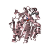
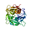
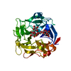
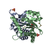
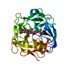
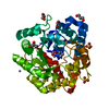
| Title | High resolution crystal structure of 1,5-alpha-arabinanase catalytic mutant (AbnBE201A) | ||||||
 Components Components | Intracellular arabinanase | ||||||
 Keywords Keywords |  HYDROLASE / Arabinanase / Glycosyl Hydrolase / High resolution / beta-propeller / Geobacillus stearothermophilus HYDROLASE / Arabinanase / Glycosyl Hydrolase / High resolution / beta-propeller / Geobacillus stearothermophilus | ||||||
| Function / homology |  Function and homology information Function and homology informationarabinan endo-1,5-alpha-L-arabinanase / arabinan endo-1,5-alpha-L-arabinosidase activity / arabinan catabolic process / metal ion binding / cytoplasmSimilarity search - Function | ||||||
| Biological species |   Geobacillus stearothermophilus (bacteria) Geobacillus stearothermophilus (bacteria) | ||||||
| Method | X-RAY DIFFRACTION / SYNCHROTRON / AB INITIO / Resolution: 1.22 Å | ||||||
 Authors Authors | Alhassid, A. / Ben David, A. / Shoham, Y. / Shoham, G. | ||||||
 Citation Citation | Journal: Biochem.J. / Year: 2009 Title: Crystal structure of an inverting GH 43 1,5-alpha-L-arabinanase from Geobacillus stearothermophilus complexed with its substrate Authors: Alhassid, A. / Ben-David, A. / Tabachnikov, O. / Libster, D. / Naveh, E. / Zolotnitsky, G. / Shoham, Y. / Shoham, G. | ||||||
| History |
|
- Structure visualization
| Structure viewer | Molecule: MolmilJmol/JSmol |
|---|
- Downloads & links
-Download
| PDBx/mmCIF format | 3d5y.cif.gz | 131.4 KB | Display | PDBx/mmCIF format |
|---|---|---|---|---|
| PDB format | pdb3d5y.ent.gz | 100.6 KB | Display | PDB format |
| PDBx/mmJSON format | 3d5y.json.gz | Tree view | PDBx/mmJSON format | |
| Others |  Other downloads Other downloads |
-Validation report
| Arichive directory | https://data.pdbj.org/pub/pdb/validation_reports/d5/3d5yftp://data.pdbj.org/pub/pdb/validation_reports/d5/3d5y | HTTPS FTP |
|---|
-Related structure data
| Related structure data | 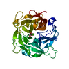 3cu9SC 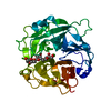 3d5zC  3d60C  3d61C S: Starting model for refinement C: citing same article ( |
|---|---|
| Similar structure data |


-Links
 PDBj
PDBj- Assembly
| Deposited unit | 
| ||||||||
|---|---|---|---|---|---|---|---|---|---|
| 1 |
| ||||||||
| 2 | 
| ||||||||
| Unit cell |
|
-Components
| #1: Protein | Mass: 35545.527 Da / Num. of mol.: 1 / Mutation: E201A Source method: isolated from a genetically manipulated source Source: (gene. exp.) Geobacillus stearothermophilus (bacteria)Strain: T-6 / Gene: abn / Plasmid: pET9d / Production host: Escherichia coli (E. coli) / Strain (production host): BL21(DE3)References: UniProt: B3EYM8, arabinan endo-1,5-alpha-L-arabinanase |
|---|---|
| #2: Chemical | ChemComp-CA /   Mass: 40.078 Da / Num. of mol.: 1 / Source method: obtained synthetically / Formula: Ca Mass: 40.078 Da / Num. of mol.: 1 / Source method: obtained synthetically / Formula: Ca |
| #3: Water | ChemComp-HOH / Water Mass: 18.015 Da / Num. of mol.: 278 / Source method: isolated from a natural source / Formula: H2O Mass: 18.015 Da / Num. of mol.: 278 / Source method: isolated from a natural source / Formula: H2O |
| Sequence details | THE 9TH RESIDUE, GLY, IS NOT A PLANNED MUTATION BUT A MUTATION IN THE CURRENT MODELS. |
-Experimental details
-Experiment
| Experiment | Method: X-RAY DIFFRACTION / Number of used crystals: 1 |
|---|
- Sample preparation
| Crystal | Density Matthews: 2.04 Å3/Da / Density % sol: 39.83 % |
|---|---|
| Crystal grow | Temperature: 295 K / Method: vapor diffusion, hanging drop / pH: 8.5 Details: 1.9M lithium sulfate, 0.1M Tris buffer pH 8.5, 4%(w/v) PEG 400, VAPOR DIFFUSION, HANGING DROP, temperature 295K |
-Data collection
| Diffraction | Mean temperature: 100 K |
|---|---|
| Diffraction source | Source: SYNCHROTRON / Site: SLS  / Beamline: X10SA / Wavelength: 0.978578 Å / Beamline: X10SA / Wavelength: 0.978578 Å |
| Detector | Type: MARMOSAIC 225 mm CCD / Detector: CCD / Date: Jun 26, 2005 |
| Radiation | Monochromator: Si 111 CHANNEL / Protocol: SINGLE WAVELENGTH / Monochromatic (M) / Laue (L): M / Scattering type: x-ray |
| Radiation wavelength | Wavelength: 0.978578 Å / Relative weight: 1 |
| Reflection | Resolution: 1.22→50 Å / Num. all: 86543 / Num. obs: 83088 / % possible obs: 96 % / Redundancy: 16 % / Biso Wilson estimate: 11.1 Å2 / Rmerge(I) obs: 0.068 / Rsym value: 0.059 / Net I/σ(I): 16.6 |
| Reflection shell | Resolution: 1.22→1.24 Å / Redundancy: 10.3 % / Rmerge(I) obs: 0.24 / Num. unique all: 3707 / Rsym value: 0.268 |
- Processing
| Software |
| |||||||||||||||||||||||||||||||||
|---|---|---|---|---|---|---|---|---|---|---|---|---|---|---|---|---|---|---|---|---|---|---|---|---|---|---|---|---|---|---|---|---|---|---|
| Refinement | Method to determine structure: AB INITIO Starting model: PDB ENTRY 3CU9 Resolution: 1.22→30 Å / Num. parameters: 21381 / Num. restraintsaints: 26021 / Isotropic thermal model: Anisotropic / Cross valid method: FREE R / σ(F): 0 / Stereochemistry target values: ENGH AND HUBER Details: ANISOTROPIC REFINEMENT REDUCED FREE R (NO CUTOFF) BY ?
| |||||||||||||||||||||||||||||||||
| Displacement parameters | Biso mean: 17.8 Å2 | |||||||||||||||||||||||||||||||||
| Refine analyze | Num. disordered residues: 0 / Occupancy sum hydrogen: 0 / Occupancy sum non hydrogen: 2797.5
| |||||||||||||||||||||||||||||||||
| Refinement step | Cycle: LAST / Resolution: 1.22→30 Å
| |||||||||||||||||||||||||||||||||
| Refine LS restraints |
| |||||||||||||||||||||||||||||||||
| LS refinement shell | Resolution: 1.22→1.3 Å / Rfactor Rfree error: 0.007
|
