 Movie
Movie Controller

[English] 日本語
 Yorodumi
- PDB-4d8l: Crystal structure of the 2-pyrone-4,6-dicarboxylic acid hydrolase... -
+ Open data

- Basic information
| Entry | Database: PDB / ID: 4d8l | |||||||||
|---|---|---|---|---|---|---|---|---|---|---|
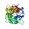
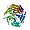
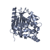
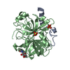
| Title | Crystal structure of the 2-pyrone-4,6-dicarboxylic acid hydrolase from sphingomonas paucimobilis | |||||||||
 Components Components | 2-pyrone-4,6-dicarbaxylate hydrolase | |||||||||
 Keywords Keywords |  HYDROLASE / STRUCTURAL GENOMICS / PROTEIN STRUCTURE INITIATIVE / NEW YORK STRUCTURAL GENOMIX RESEARCH CONSORTIUM / NYSGXRC / PSI-2 / New York SGX Research Center for Structural Genomics HYDROLASE / STRUCTURAL GENOMICS / PROTEIN STRUCTURE INITIATIVE / NEW YORK STRUCTURAL GENOMIX RESEARCH CONSORTIUM / NYSGXRC / PSI-2 / New York SGX Research Center for Structural Genomics | |||||||||
| Function / homology |  Function and homology information2-pyrone-4,6-dicarboxylate lactonase / 2-pyrone-4,6-dicarboxylate lactonase activity / 3,4-dihydroxybenzoate catabolic process / lignin catabolic process Function and homology information2-pyrone-4,6-dicarboxylate lactonase / 2-pyrone-4,6-dicarboxylate lactonase activity / 3,4-dihydroxybenzoate catabolic process / lignin catabolic processSimilarity search - Function | |||||||||
| Biological species |  Sphingomonas paucimobilis (bacteria) Sphingomonas paucimobilis (bacteria) | |||||||||
| Method | X-RAY DIFFRACTION / SYNCHROTRON / SAD / Resolution: 2 Å | |||||||||
 Authors Authors | Malashkevich, V.N. / Toro, R. / Bonanno, J. / Sauder, J.M. / Burley, S.K. / Almo, S.C. / New York SGX Research Center for Structural Genomics (NYSGXRC) | |||||||||
 Citation Citation | Journal: Biochemistry / Year: 2012 Title: Structure and Catalytic Mechanism of LigI: Insight into the Amidohydrolase Enzymes of cog3618 and Lignin Degradation. Authors: Hobbs, M.E. / Malashkevich, V. / Williams, H.J. / Xu, C. / Sauder, J.M. / Burley, S.K. / Almo, S.C. / Raushel, F.M. | |||||||||
| History |
|
- Structure visualization
| Structure viewer | Molecule: MolmilJmol/JSmol |
|---|
- Downloads & links
-Download
| PDBx/mmCIF format | 4d8l.cif.gz | 131.2 KB | Display | PDBx/mmCIF format |
|---|---|---|---|---|
| PDB format | pdb4d8l.ent.gz | 103 KB | Display | PDB format |
| PDBx/mmJSON format | 4d8l.json.gz | Tree view | PDBx/mmJSON format | |
| Others |  Other downloads Other downloads |
-Validation report
| Arichive directory | https://data.pdbj.org/pub/pdb/validation_reports/d8/4d8lftp://data.pdbj.org/pub/pdb/validation_reports/d8/4d8l | HTTPS FTP |
|---|
-Related structure data
| Related structure data | 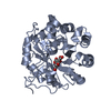 4di8C 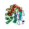 4di9C 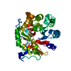 4diaC C: citing same article ( |
|---|---|
| Similar structure data | |
| Other databases |


-Links
 PDBj
PDBj

- Assembly
| Deposited unit | 
| ||||||||
|---|---|---|---|---|---|---|---|---|---|
| 1 |
| ||||||||
| Unit cell |
| ||||||||
| Details | monomer |
-Components
| #1: Protein | Mass: 34030.770 Da / Num. of mol.: 1 Source method: isolated from a genetically manipulated source Source: (gene. exp.) Sphingomonas paucimobilis (bacteria) / Strain: SYK-6 / Gene: ligI, SLG_12570 / Plasmid: BC-PSGX3(BC) / Production host: Escherichia coli (E. coli) / Strain (production host): BL21(DE3)References: UniProt: O87170, 2-pyrone-4,6-dicarboxylate lactonase |
|---|---|
| #2: Water | ChemComp-HOH / Water Mass: 18.015 Da / Num. of mol.: 235 / Source method: isolated from a natural source / Formula: H2O Mass: 18.015 Da / Num. of mol.: 235 / Source method: isolated from a natural source / Formula: H2O |
-Experimental details
-Experiment
| Experiment | Method: X-RAY DIFFRACTION / Number of used crystals: 1 |
|---|
- Sample preparation
| Crystal | Density Matthews: 2.32 Å3/Da / Density % sol: 46.92 % |
|---|---|
| Crystal grow | Temperature: 298 K / Method: vapor diffusion, sitting drop / pH: 8.5 Details: 30% PEG 4000, 0.1M TRIS-HCL PH 8.5,0.2 MG CHLORIDE, VAPOR DIFFUSION, SITTING DROP, temperature 298K |
-Data collection
| Diffraction | Mean temperature: 100 K | |||||||||||||||||||||||||||||||||||||||||||||||||||||||||||||||||||||||||||||
|---|---|---|---|---|---|---|---|---|---|---|---|---|---|---|---|---|---|---|---|---|---|---|---|---|---|---|---|---|---|---|---|---|---|---|---|---|---|---|---|---|---|---|---|---|---|---|---|---|---|---|---|---|---|---|---|---|---|---|---|---|---|---|---|---|---|---|---|---|---|---|---|---|---|---|---|---|---|---|
| Diffraction source | Source: SYNCHROTRON / Site: NSLS  / Beamline: X29A / Wavelength: 0.9791 Å / Beamline: X29A / Wavelength: 0.9791 Å | |||||||||||||||||||||||||||||||||||||||||||||||||||||||||||||||||||||||||||||
| Detector | Type: ADSC QUANTUM 315 / Detector: CCD / Date: Apr 27, 2007 | |||||||||||||||||||||||||||||||||||||||||||||||||||||||||||||||||||||||||||||
| Radiation | Protocol: SINGLE WAVELENGTH / Scattering type: x-ray | |||||||||||||||||||||||||||||||||||||||||||||||||||||||||||||||||||||||||||||
| Radiation wavelength | Wavelength: 0.9791 Å / Relative weight: 1 | |||||||||||||||||||||||||||||||||||||||||||||||||||||||||||||||||||||||||||||
| Reflection | Redundancy: 1.8 % / Av σ(I) over netI: 16.69 / Number: 61399 / Rmerge(I) obs: 0.057 / Χ2: 1.61 / D res high: 2 Å / D res low: 50 Å / Num. obs: 35073 / % possible obs: 84.2 | |||||||||||||||||||||||||||||||||||||||||||||||||||||||||||||||||||||||||||||
| Diffraction reflection shell |
| |||||||||||||||||||||||||||||||||||||||||||||||||||||||||||||||||||||||||||||
| Reflection | Resolution: 2→50 Å / Num. obs: 35073 / % possible obs: 84.2 % / Redundancy: 1.8 % / Rmerge(I) obs: 0.057 / Χ2: 1.608 / Net I/σ(I): 11.3 | |||||||||||||||||||||||||||||||||||||||||||||||||||||||||||||||||||||||||||||
| Reflection shell |
|
-Phasing
| Phasing | Method: SAD |
|---|
- Processing
| Software |
| |||||||||||||||||||||||||||||||||||||||||||||
|---|---|---|---|---|---|---|---|---|---|---|---|---|---|---|---|---|---|---|---|---|---|---|---|---|---|---|---|---|---|---|---|---|---|---|---|---|---|---|---|---|---|---|---|---|---|---|
| Refinement | Method to determine structure: SAD / Resolution: 2→19.91 Å / Cor.coef. Fo:Fc: 0.962 / Cor.coef. Fo:Fc free: 0.927 / WRfactor Rfree: 0.21 / WRfactor Rwork: 0.1576 / Occupancy max: 1 / Occupancy min: 1 / FOM work R set: 0.8348 / SU B: 8.297 / SU ML: 0.123 / SU R Cruickshank DPI: 0.2489 / SU Rfree: 0.2015 / Cross valid method: THROUGHOUT / σ(F): 0 / ESU R: 0.249 / ESU R Free: 0.202 / Stereochemistry target values: MAXIMUM LIKELIHOOD Details: U VALUES : WITH TLS ADDED HYDROGENS HAVE BEEN USED IF PRESENT IN THE INPUT
| |||||||||||||||||||||||||||||||||||||||||||||
| Solvent computation | Ion probe radii: 0.8 Å / Shrinkage radii: 0.8 Å / VDW probe radii: 1.2 Å / Solvent model: MASK | |||||||||||||||||||||||||||||||||||||||||||||
| Displacement parameters | Biso max: 84.76 Å2 / Biso mean: 33.881 Å2 / Biso min: 12.96 Å2
| |||||||||||||||||||||||||||||||||||||||||||||
| Refinement step | Cycle: LAST / Resolution: 2→19.91 Å
| |||||||||||||||||||||||||||||||||||||||||||||
| Refine LS restraints |
| |||||||||||||||||||||||||||||||||||||||||||||
| LS refinement shell | Resolution: 2.002→2.054 Å / Total num. of bins used: 20
| |||||||||||||||||||||||||||||||||||||||||||||
| Refinement TLS params. | Method: refined / Origin x: 40.7749 Å / Origin y: 39.4309 Å / Origin z: 9.6797 Å
|
