 Movie
Movie Controller

[English] 日本語
 Yorodumi
- PDB-3czl: Crystal Structure Analysis of Sucrose hydrolase(SUH) E322Q-glucos... -
+ Open data

- Basic information
| Entry | Database: PDB / ID: 3czl | ||||||
|---|---|---|---|---|---|---|---|
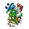
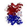
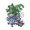
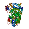
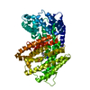
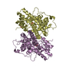
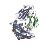
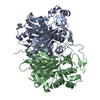
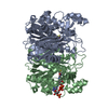
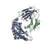
| Title | Crystal Structure Analysis of Sucrose hydrolase(SUH) E322Q-glucose complex | ||||||
 Components Components | sucrose hydrolase | ||||||
 Keywords Keywords |  HYDROLASE / (alpha/beta)8-barrel HYDROLASE / (alpha/beta)8-barrel | ||||||
| Function / homology |  Function and homology informationamylosucrase activity / hydrolase activity / carbohydrate metabolic process Function and homology informationamylosucrase activity / hydrolase activity / carbohydrate metabolic processSimilarity search - Function | ||||||
| Biological species |  Xanthomonas axonopodis pv. glycines (bacteria) Xanthomonas axonopodis pv. glycines (bacteria) | ||||||
| Method | X-RAY DIFFRACTION / SYNCHROTRON / MOLECULAR REPLACEMENT / Resolution: 2 Å | ||||||
 Authors Authors | Kim, M.I. / Rhee, S. | ||||||
 Citation Citation | Journal: J.Mol.Biol. / Year: 2008 Title: Crystal structures and mutagenesis of sucrose hydrolase from Xanthomonas axonopodis pv. glycines: insight into the exclusively hydrolytic amylosucrase fold. Authors: Kim, M.I. / Kim, H.S. / Jung, J. / Rhee, S. | ||||||
| History |
|
- Structure visualization
| Structure viewer | Molecule: MolmilJmol/JSmol |
|---|
- Downloads & links
-Download
| PDBx/mmCIF format | 3czl.cif.gz | 139.7 KB | Display | PDBx/mmCIF format |
|---|---|---|---|---|
| PDB format | pdb3czl.ent.gz | 107.4 KB | Display | PDB format |
| PDBx/mmJSON format | 3czl.json.gz | Tree view | PDBx/mmJSON format | |
| Others |  Other downloads Other downloads |
-Validation report
| Arichive directory | https://data.pdbj.org/pub/pdb/validation_reports/cz/3czlftp://data.pdbj.org/pub/pdb/validation_reports/cz/3czl | HTTPS FTP |
|---|
-Related structure data
| Related structure data |  3czeSC  3czgC 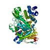 3czkC S: Starting model for refinement C: citing same article ( |
|---|---|
| Similar structure data |
-Links
 PDBj
PDBj


- Assembly
| Deposited unit | 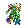
| ||||||||
|---|---|---|---|---|---|---|---|---|---|
| 1 |
| ||||||||
| Unit cell |
|
-Components
| #1: Protein | Mass: 70420.070 Da / Num. of mol.: 1 / Mutation: E322Q Source method: isolated from a genetically manipulated source Source: (gene. exp.) Xanthomonas axonopodis pv. glycines (bacteria)Strain: 8ra / Gene: supH / Plasmid: pET41b / Production host: Escherichia coli (E. coli) / Strain (production host): B834(DE3)pLysS / References: UniProt: Q6UVM5, sucrose alpha-glucosidase |
|---|---|
| #2: Sugar | ChemComp-GLC / Glucose  Type: D-saccharide, alpha linking / Mass: 180.156 Da / Num. of mol.: 1 Type: D-saccharide, alpha linking / Mass: 180.156 Da / Num. of mol.: 1Source method: isolated from a genetically manipulated source Formula: C6H12O6 |
| #3: Water | ChemComp-HOH / Water Mass: 18.015 Da / Num. of mol.: 398 / Source method: isolated from a natural source / Formula: H2O Mass: 18.015 Da / Num. of mol.: 398 / Source method: isolated from a natural source / Formula: H2O |
-Experimental details
-Experiment
| Experiment | Method: X-RAY DIFFRACTION / Number of used crystals: 1 |
|---|
- Sample preparation
| Crystal | Density Matthews: 2.28 Å3/Da / Density % sol: 45.95 % / Mosaicity: 0.334 ° |
|---|---|
| Crystal grow | Temperature: 295 K / Method: vapor diffusion, hanging drop / pH: 6.5 Details: 0.1M HEPES, 0.2M calcium acetate, 16% PEG 3000, 5mM DTT, 0.1M cesium chloride , pH 6.5, VAPOR DIFFUSION, HANGING DROP, temperature 295K |
-Data collection
| Diffraction | Mean temperature: 100 K |
|---|---|
| Diffraction source | Source: SYNCHROTRON / Site: PAL/PLS  / Beamline: 6C1 / Wavelength: 1.2399 Å / Beamline: 6C1 / Wavelength: 1.2399 Å |
| Detector | Type: ADSC QUANTUM 210 / Detector: CCD / Date: Feb 23, 2007 |
| Radiation | Protocol: SINGLE WAVELENGTH / Monochromatic (M) / Laue (L): M / Scattering type: x-ray |
| Radiation wavelength | Wavelength: 1.2399 Å / Relative weight: 1 |
| Reflection | Resolution: 2→50 Å / Num. obs: 44083 / % possible obs: 99 % / Redundancy: 6.8 % / Rmerge(I) obs: 0.054 / Χ2: 1.17 / Net I/σ(I): 16.1 |
| Reflection shell | Resolution: 2→2.07 Å / Redundancy: 5.2 % / Rmerge(I) obs: 0.143 / Num. unique all: 4001 / Χ2: 0.581 / % possible all: 91.2 |
- Processing
| Software |
| ||||||||||||||||||||||||||||||||||||
|---|---|---|---|---|---|---|---|---|---|---|---|---|---|---|---|---|---|---|---|---|---|---|---|---|---|---|---|---|---|---|---|---|---|---|---|---|---|
| Refinement | Method to determine structure: MOLECULAR REPLACEMENT Starting model: 3CZE Resolution: 2→50 Å
| ||||||||||||||||||||||||||||||||||||
| Solvent computation | Bsol: 51.731 Å2 | ||||||||||||||||||||||||||||||||||||
| Displacement parameters | Biso mean: 28.778 Å2
| ||||||||||||||||||||||||||||||||||||
| Refinement step | Cycle: LAST / Resolution: 2→50 Å
| ||||||||||||||||||||||||||||||||||||
| Refine LS restraints |
| ||||||||||||||||||||||||||||||||||||
| Xplor file |
|
