 Movie
Movie Controller

[English] 日本語
 Yorodumi
- PDB-3wj8: Crystal Structure of DL-2-haloacid dehalogenase mutant with 2-bro... -
+ Open data

- Basic information
| Entry | Database: PDB / ID: 3wj8 | ||||||
|---|---|---|---|---|---|---|---|
| Title | Crystal Structure of DL-2-haloacid dehalogenase mutant with 2-bromo-2-methylpropionate | ||||||
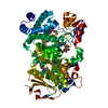
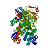
 Components Components | DL-2-haloacid dehalogenase | ||||||
 Keywords Keywords |  HYDROLASE / dehalogenases HYDROLASE / dehalogenases | ||||||
| Function / homology | 2-haloacid dehalogenase (configuration-inverting) / DL-2 haloacid dehalogenase activity / 2-bromo-2-methylpropanoic acid / DL-2-haloacid dehalogenase Function and homology information Function and homology information | ||||||
| Biological species |  Methylobacterium (bacteria) Methylobacterium (bacteria) | ||||||
| Method | X-RAY DIFFRACTION / MOLECULAR REPLACEMENT / Resolution: 2.7 Å | ||||||
 Authors Authors | Siwek, A. / Omi, R. / Hirotsu, K. / Jitsumori, K. / Esaki, N. / Kurihara, T. / Paneth, P. | ||||||
 Citation Citation | Journal: Arch.Biochem.Biophys. / Year: 2013 Title: Binding modes of DL-2-haloacid dehalogenase revealed by crystallography, modeling and isotope effects studies. Authors: Siwek, A. / Omi, R. / Hirotsu, K. / Jitsumori, K. / Esaki, N. / Kurihara, T. / Paneth, P. | ||||||
| History |
|
- Structure visualization
| Structure viewer | Molecule: MolmilJmol/JSmol |
|---|
- Downloads & links
-Download
| PDBx/mmCIF format | 3wj8.cif.gz | 361.1 KB | Display | PDBx/mmCIF format |
|---|---|---|---|---|
| PDB format | pdb3wj8.ent.gz | 299.5 KB | Display | PDB format |
| PDBx/mmJSON format | 3wj8.json.gz | Tree view | PDBx/mmJSON format | |
| Others |  Other downloads Other downloads |
-Validation report
| Arichive directory | https://data.pdbj.org/pub/pdb/validation_reports/wj/3wj8ftp://data.pdbj.org/pub/pdb/validation_reports/wj/3wj8 | HTTPS FTP |
|---|
-Related structure data


-Links
 PDBj
PDBj
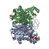
- Assembly
| Deposited unit | 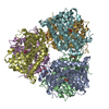
| ||||||||
|---|---|---|---|---|---|---|---|---|---|
| 1 | 
| ||||||||
| 2 | 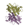
| ||||||||
| 3 | 
| ||||||||
| Unit cell |
|
-Components
| #1: Protein | Mass: 34092.922 Da / Num. of mol.: 6 / Mutation: D54H, D194N Source method: isolated from a genetically manipulated source Source: (gene. exp.) Methylobacterium (bacteria) / Genus: dl-dex / Strain: CPA1Carboxypeptidase A1 / Production host: Escherichia coli (E. coli)References: UniProt: A6BM74, 2-haloacid dehalogenase (configuration-inverting)#2: Chemical | ChemComp-B2P /   Mass: 167.001 Da / Num. of mol.: 6 / Source method: obtained synthetically / Formula: C4H7BrO2 Mass: 167.001 Da / Num. of mol.: 6 / Source method: obtained synthetically / Formula: C4H7BrO2#3: Chemical | ChemComp-GOL / Glycerol  Mass: 92.094 Da / Num. of mol.: 6 / Source method: obtained synthetically / Formula: C3H8O3 Mass: 92.094 Da / Num. of mol.: 6 / Source method: obtained synthetically / Formula: C3H8O3#4: Water | ChemComp-HOH / | Water Mass: 18.015 Da / Num. of mol.: 611 / Source method: isolated from a natural source / Formula: H2O Mass: 18.015 Da / Num. of mol.: 611 / Source method: isolated from a natural source / Formula: H2O |
|---|
-Experimental details
-Experiment
| Experiment | Method: X-RAY DIFFRACTION |
|---|
- Sample preparation
| Crystal | Density Matthews: 2.8 Å3/Da / Density % sol: 56.07 % |
|---|---|
| Crystal grow | Temperature: 298 K / Method: vapor diffusion / pH: 7.5 / Details: pH 7.5, VAPOR DIFFUSION, temperature 298K |
-Data collection
| Diffraction source | Source: ROTATING ANODE / Type: RIGAKU / Wavelength: 1.5418 Å |
|---|---|
| Radiation | Protocol: SINGLE WAVELENGTH / Monochromatic (M) / Laue (L): M / Scattering type: x-ray |
| Radiation wavelength | Wavelength: 1.5418 Å / Relative weight: 1 |
| Reflection | Resolution: 2.7→161.35 Å / Num. obs: 61648 / % possible obs: 99.5 % |
- Processing
| Software | Name: REFMAC / Version: 5.6.0117 / Classification: refinement | ||||||||||||||||||||||||||||||||||||||||||||||||||||||||||||||||||||||||||||||||||||||||||||||||||||||||||||||||||||||||||||||||||||||||||||||||||||||||||||||||||||||||||||||||||||||
|---|---|---|---|---|---|---|---|---|---|---|---|---|---|---|---|---|---|---|---|---|---|---|---|---|---|---|---|---|---|---|---|---|---|---|---|---|---|---|---|---|---|---|---|---|---|---|---|---|---|---|---|---|---|---|---|---|---|---|---|---|---|---|---|---|---|---|---|---|---|---|---|---|---|---|---|---|---|---|---|---|---|---|---|---|---|---|---|---|---|---|---|---|---|---|---|---|---|---|---|---|---|---|---|---|---|---|---|---|---|---|---|---|---|---|---|---|---|---|---|---|---|---|---|---|---|---|---|---|---|---|---|---|---|---|---|---|---|---|---|---|---|---|---|---|---|---|---|---|---|---|---|---|---|---|---|---|---|---|---|---|---|---|---|---|---|---|---|---|---|---|---|---|---|---|---|---|---|---|---|---|---|---|---|
| Refinement | Method to determine structure: MOLECULAR REPLACEMENT / Resolution: 2.7→50 Å / Cor.coef. Fo:Fc: 0.933 / Cor.coef. Fo:Fc free: 0.86 / SU B: 13.519 / SU ML: 0.271 / Cross valid method: THROUGHOUT / ESU R Free: 0.392 / Stereochemistry target values: MAXIMUM LIKELIHOOD / Details: HYDROGENS HAVE BEEN USED IF PRESENT IN THE INPUT
| ||||||||||||||||||||||||||||||||||||||||||||||||||||||||||||||||||||||||||||||||||||||||||||||||||||||||||||||||||||||||||||||||||||||||||||||||||||||||||||||||||||||||||||||||||||||
| Solvent computation | Ion probe radii: 0.8 Å / Shrinkage radii: 0.8 Å / VDW probe radii: 1.2 Å / Solvent model: MASK | ||||||||||||||||||||||||||||||||||||||||||||||||||||||||||||||||||||||||||||||||||||||||||||||||||||||||||||||||||||||||||||||||||||||||||||||||||||||||||||||||||||||||||||||||||||||
| Displacement parameters | Biso mean: 16.812 Å2
| ||||||||||||||||||||||||||||||||||||||||||||||||||||||||||||||||||||||||||||||||||||||||||||||||||||||||||||||||||||||||||||||||||||||||||||||||||||||||||||||||||||||||||||||||||||||
| Refinement step | Cycle: LAST / Resolution: 2.7→50 Å
| ||||||||||||||||||||||||||||||||||||||||||||||||||||||||||||||||||||||||||||||||||||||||||||||||||||||||||||||||||||||||||||||||||||||||||||||||||||||||||||||||||||||||||||||||||||||
| Refine LS restraints |
|