 Movie
Movie Controller

[English] 日本語
 Yorodumi
- PDB-3cuk: Crystal structure of human D-amino acid oxidase: bound to an inhibitor -
+ Open data

- Basic information
| Entry | Database: PDB / ID: 3cuk | ||||||
|---|---|---|---|---|---|---|---|
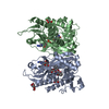
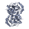
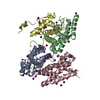
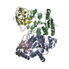
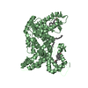
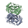
| Title | Crystal structure of human D-amino acid oxidase: bound to an inhibitor | ||||||
 Components Components | D-amino-acid oxidase D-amino acid oxidase D-amino acid oxidase | ||||||
 Keywords Keywords | OXIDOREDUCTASE / ALPHA-BETA-ALPHA MOTIF / FLAVIN CONTAINING PROTEINALPHA-BETA-ALPHA MOTIF / FLAVIN CONTAINING PROTEIN / FAD / Flavoprotein / Peroxisome | ||||||
| Function / homology |  Function and homology information Function and homology informationD-alanine catabolic process / D-amino-acid oxidase / D-amino-acid oxidase activity / D-serine metabolic process / proline catabolic process / D-amino acid catabolic process / D-serine catabolic process / Glyoxylate metabolism and glycine degradation / dopamine biosynthetic process / presynaptic active zone ...D-alanine catabolic process / D-amino-acid oxidase / D-amino-acid oxidase activity / D-serine metabolic process / proline catabolic process / D-amino acid catabolic process / D-serine catabolic process / Glyoxylate metabolism and glycine degradation / dopamine biosynthetic process / presynaptic active zone / neutrophil-mediated killing of gram-negative bacterium / peroxisomal matrix / digestion / FAD binding / Peroxisomal protein import / identical protein binding / cytosol / cytoplasmSimilarity search - Function | ||||||
| Biological species |  Homo sapiens (human) Homo sapiens (human) | ||||||
| Method | X-RAY DIFFRACTION / SYNCHROTRON / MOLECULAR REPLACEMENT / Resolution: 2.49 Å | ||||||
 Authors Authors | Prasad, S. / Munshi, S. | ||||||
 Citation Citation | Journal: Bioorg.Med.Chem.Lett. / Year: 2008 Title: The discovery of fused pyrrole carboxylic acids as novel, potent D-amino acid oxidase (DAO) inhibitors. Authors: Sparey, T. / Abeywickrema, P. / Almond, S. / Brandon, N. / Byrne, N. / Campbell, A. / Hutson, P.H. / Jacobson, M. / Jones, B. / Munshi, S. / Pascarella, D. / Pike, A. / Prasad, G.S. / Sachs, ...Authors: Sparey, T. / Abeywickrema, P. / Almond, S. / Brandon, N. / Byrne, N. / Campbell, A. / Hutson, P.H. / Jacobson, M. / Jones, B. / Munshi, S. / Pascarella, D. / Pike, A. / Prasad, G.S. / Sachs, N. / Sakatis, M. / Sardana, V. / Venkatraman, S. / Young, M.B. | ||||||
| History |
|
- Structure visualization
| Structure viewer | Molecule: MolmilJmol/JSmol |
|---|
- Downloads & links
-Download
| PDBx/mmCIF format | 3cuk.cif.gz | 276.2 KB | Display | PDBx/mmCIF format |
|---|---|---|---|---|
| PDB format | pdb3cuk.ent.gz | 222.7 KB | Display | PDB format |
| PDBx/mmJSON format | 3cuk.json.gz | Tree view | PDBx/mmJSON format | |
| Others |  Other downloads Other downloads |
-Validation report
| Arichive directory | https://data.pdbj.org/pub/pdb/validation_reports/cu/3cukftp://data.pdbj.org/pub/pdb/validation_reports/cu/3cuk | HTTPS FTP |
|---|
-Related structure data
| Related structure data | 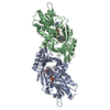 1ve9S S: Starting model for refinement |
|---|---|
| Similar structure data |


-Links
 PDBj
PDBj- Assembly
| Deposited unit | 
| ||||||||
|---|---|---|---|---|---|---|---|---|---|
| 1 | 
| ||||||||
| 2 | 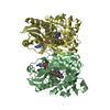
| ||||||||
| 3 | 
| ||||||||
| 4 | 
| ||||||||
| Unit cell |
|
-Components
| #1: Protein | D-amino acid oxidase / DAMOX / DAO / DAAO Mass: 39520.910 Da / Num. of mol.: 4 Source method: isolated from a genetically manipulated source Source: (gene. exp.) Homo sapiens (human) / Gene: DAO, DAMOX / Plasmid: pRSET / Production host:  Escherichia coli (E. coli) / Strain (production host): BL21(DE3) / References: UniProt: P14920, D-amino-acid oxidase Escherichia coli (E. coli) / Strain (production host): BL21(DE3) / References: UniProt: P14920, D-amino-acid oxidase#2: Chemical | ChemComp-FAD / Flavin adenine dinucleotide  Mass: 785.550 Da / Num. of mol.: 4 / Source method: obtained synthetically / Formula: C27H33N9O15P2 / Comment: FAD*YM Mass: 785.550 Da / Num. of mol.: 4 / Source method: obtained synthetically / Formula: C27H33N9O15P2 / Comment: FAD*YM#3: Chemical | ChemComp-4P5 /   Mass: 151.119 Da / Num. of mol.: 4 / Source method: obtained synthetically / Formula: C7H5NO3 Mass: 151.119 Da / Num. of mol.: 4 / Source method: obtained synthetically / Formula: C7H5NO3#4: Water | ChemComp-HOH / | Water Mass: 18.015 Da / Num. of mol.: 42 / Source method: isolated from a natural source / Formula: H2O Mass: 18.015 Da / Num. of mol.: 42 / Source method: isolated from a natural source / Formula: H2O |
|---|
-Experimental details
-Experiment
| Experiment | Method: X-RAY DIFFRACTION / Number of used crystals: 1 |
|---|
- Sample preparation
| Crystal | Density Matthews: 2.21 Å3/Da / Density % sol: 44.38 % |
|---|---|
| Crystal grow | Temperature: 298 K / Method: vapor diffusion, sitting drop / pH: 7.8 Details: 15% PEG 3350, 0.15M POTASSIUM TRICITR TRIS-HCL, pH 7.80, VAPOR DIFFUSION, SITTING DROP, temperature 298K |
-Data collection
| Diffraction | Mean temperature: 100 K |
|---|---|
| Diffraction source | Source: SYNCHROTRON / Wavelength: 1 |
| Detector | Detector: IMAGE PLATE / Date: Feb 20, 2005 |
| Radiation | Protocol: SINGLE WAVELENGTH / Monochromatic (M) / Laue (L): M / Scattering type: x-ray |
| Radiation wavelength | Wavelength: 1 Å / Relative weight: 1 |
| Reflection | Resolution: 2.49→50 Å / Num. obs: 46710 / % possible obs: 96.8 % / Redundancy: 2.9 % / Rmerge(I) obs: 0.097 / Net I/σ(I): 11 |
| Reflection shell | Resolution: 2.49→2.59 Å / Redundancy: 2.9 % / Rmerge(I) obs: 0.558 / Mean I/σ(I) obs: 2 / % possible all: 95.4 |
- Processing
| Software |
| ||||||||||||||||||||||||||||||||||||||||||||||||||||||||||||||||||||||||||||||||||||||||||||||||||||||||||||||||||||||||||||||||||||||||||||||||||||||||||||||||||||||||||
|---|---|---|---|---|---|---|---|---|---|---|---|---|---|---|---|---|---|---|---|---|---|---|---|---|---|---|---|---|---|---|---|---|---|---|---|---|---|---|---|---|---|---|---|---|---|---|---|---|---|---|---|---|---|---|---|---|---|---|---|---|---|---|---|---|---|---|---|---|---|---|---|---|---|---|---|---|---|---|---|---|---|---|---|---|---|---|---|---|---|---|---|---|---|---|---|---|---|---|---|---|---|---|---|---|---|---|---|---|---|---|---|---|---|---|---|---|---|---|---|---|---|---|---|---|---|---|---|---|---|---|---|---|---|---|---|---|---|---|---|---|---|---|---|---|---|---|---|---|---|---|---|---|---|---|---|---|---|---|---|---|---|---|---|---|---|---|---|---|---|---|---|
| Refinement | Method to determine structure: MOLECULAR REPLACEMENT Starting model: PDB ENTRY 1VE9 Resolution: 2.49→42.52 Å / Cor.coef. Fo:Fc: 0.943 / Cor.coef. Fo:Fc free: 0.888 / SU B: 29.033 / SU ML: 0.317 / TLS residual ADP flag: LIKELY RESIDUAL / Cross valid method: THROUGHOUT / ESU R: 2.129 / ESU R Free: 0.417 / Stereochemistry target values: MAXIMUM LIKELIHOOD / Details: HYDROGENS HAVE BEEN ADDED IN THE RIDING POSITIONS
| ||||||||||||||||||||||||||||||||||||||||||||||||||||||||||||||||||||||||||||||||||||||||||||||||||||||||||||||||||||||||||||||||||||||||||||||||||||||||||||||||||||||||||
| Solvent computation | Ion probe radii: 0.8 Å / Shrinkage radii: 0.8 Å / VDW probe radii: 1.2 Å / Solvent model: MASK | ||||||||||||||||||||||||||||||||||||||||||||||||||||||||||||||||||||||||||||||||||||||||||||||||||||||||||||||||||||||||||||||||||||||||||||||||||||||||||||||||||||||||||
| Displacement parameters | Biso mean: 23.324 Å2
| ||||||||||||||||||||||||||||||||||||||||||||||||||||||||||||||||||||||||||||||||||||||||||||||||||||||||||||||||||||||||||||||||||||||||||||||||||||||||||||||||||||||||||
| Refinement step | Cycle: LAST / Resolution: 2.49→42.52 Å
| ||||||||||||||||||||||||||||||||||||||||||||||||||||||||||||||||||||||||||||||||||||||||||||||||||||||||||||||||||||||||||||||||||||||||||||||||||||||||||||||||||||||||||
| Refine LS restraints |
| ||||||||||||||||||||||||||||||||||||||||||||||||||||||||||||||||||||||||||||||||||||||||||||||||||||||||||||||||||||||||||||||||||||||||||||||||||||||||||||||||||||||||||
| LS refinement shell | Resolution: 2.492→2.557 Å / Total num. of bins used: 20
| ||||||||||||||||||||||||||||||||||||||||||||||||||||||||||||||||||||||||||||||||||||||||||||||||||||||||||||||||||||||||||||||||||||||||||||||||||||||||||||||||||||||||||
| Refinement TLS params. | Method: refined / Refine-ID: X-RAY DIFFRACTION
| ||||||||||||||||||||||||||||||||||||||||||||||||||||||||||||||||||||||||||||||||||||||||||||||||||||||||||||||||||||||||||||||||||||||||||||||||||||||||||||||||||||||||||
| Refinement TLS group |
|
