 Movie
Movie Controller

[English] 日本語
 Yorodumi
- PDB-3bor: Crystal structure of the DEADc domain of human translation initia... -
+ Open data

- Basic information
| Entry | Database: PDB / ID: 3bor | ||||||
|---|---|---|---|---|---|---|---|
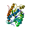
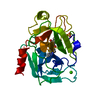
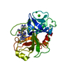
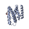
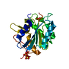
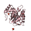
| Title | Crystal structure of the DEADc domain of human translation initiation factor 4A-2 | ||||||
 Components Components | Human initiation factor 4A-II | ||||||
 Keywords Keywords |  HYDROLASE / translation initiation / DEAD box / structural genomics / helicase / ATP-binding / Host-virus interaction / Initiation factor / Nucleotide-binding / Protein biosynthesis / RNA-binding / Structural Genomics Consortium / SGC HYDROLASE / translation initiation / DEAD box / structural genomics / helicase / ATP-binding / Host-virus interaction / Initiation factor / Nucleotide-binding / Protein biosynthesis / RNA-binding / Structural Genomics Consortium / SGC | ||||||
| Function / homology |  Function and homology information Function and homology informationnegative regulation of RNA-dependent RNA polymerase activity / Activation of the mRNA upon binding of the cap-binding complex and eIFs, and subsequent binding to 43S / eukaryotic translation initiation factor 4F complex / cytoplasmic translational initiation / Deadenylation of mRNA / regulation of translational initiation / Ribosomal scanning and start codon recognition / Translation initiation complex formation / GTP hydrolysis and joining of the 60S ribosomal subunit / L13a-mediated translational silencing of Ceruloplasmin expression ...negative regulation of RNA-dependent RNA polymerase activity / Activation of the mRNA upon binding of the cap-binding complex and eIFs, and subsequent binding to 43S / eukaryotic translation initiation factor 4F complex / cytoplasmic translational initiation / Deadenylation of mRNA / regulation of translational initiation / Ribosomal scanning and start codon recognition / Translation initiation complex formation / GTP hydrolysis and joining of the 60S ribosomal subunit / L13a-mediated translational silencing of Ceruloplasmin expression / translational initiation / translation initiation factor activity / viral process / helicase activity / cellular response to leukemia inhibitory factor / ISG15 antiviral mechanism / RNA helicase activity / RNA helicase / perinuclear region of cytoplasm / ATP hydrolysis activity / RNA binding / ATP binding / cytosolSimilarity search - Function | ||||||
| Biological species |  Homo sapiens (human) Homo sapiens (human) | ||||||
| Method | X-RAY DIFFRACTION / SYNCHROTRON / MOLECULAR REPLACEMENT / molecular replacement / Resolution: 1.85 Å | ||||||
 Authors Authors | Dimov, S. / Hong, B. / Tempel, W. / MacKenzie, F. / Karlberg, T. / Arrowsmith, C.H. / Edwards, A.M. / Weigelt, J. / Bochkarev, A. / Park, H. / Structural Genomics Consortium (SGC) | ||||||
 Citation Citation | Journal: Plos One / Year: 2010 Title: Comparative Structural Analysis of Human DEAD-Box RNA Helicases. Authors: Schutz, P. / Karlberg, T. / van den Berg, S. / Collins, R. / Lehtio, L. / Hogbom, M. / Holmberg-Schiavone, L. / Tempel, W. / Park, H.W. / Hammarstrom, M. / Moche, M. / Thorsell, A.G. / Schuler, H. | ||||||
| History |
|
- Structure visualization
| Structure viewer | Molecule: MolmilJmol/JSmol |
|---|
- Downloads & links
-Download
| PDBx/mmCIF format | 3bor.cif.gz | 54.8 KB | Display | PDBx/mmCIF format |
|---|---|---|---|---|
| PDB format | pdb3bor.ent.gz | 37.6 KB | Display | PDB format |
| PDBx/mmJSON format | 3bor.json.gz | Tree view | PDBx/mmJSON format | |
| Others |  Other downloads Other downloads |
-Validation report
| Arichive directory | https://data.pdbj.org/pub/pdb/validation_reports/bo/3borftp://data.pdbj.org/pub/pdb/validation_reports/bo/3bor | HTTPS FTP |
|---|
-Related structure data
| Related structure data | 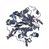 2g9nSC 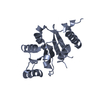 2p6nC  2pl3C  2rb4C 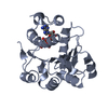 3b7gC 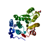 3berC 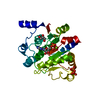 3dkpC  3fe2C  3iuyC  3ly5C S: Starting model for refinement C: citing same article ( |
|---|---|
| Similar structure data |


-Links
 PDBj
PDBj


- Assembly
| Deposited unit | 
| ||||||||
|---|---|---|---|---|---|---|---|---|---|
| 1 |
| ||||||||
| Unit cell |
| ||||||||
| Details | AUTHORS STATE THAT THE BIOLOGICAL UNIT OF THIS PROTEIN IS UNKNOWN. |
-Components
| #1: Protein | Mass: 26927.096 Da / Num. of mol.: 1 / Fragment: DEADc domain: Residues 22-240 Source method: isolated from a genetically manipulated source Source: (gene. exp.) Homo sapiens (human) / Gene: EIF4A2, DDX2B, EIF4F / Plasmid: pET28-mhl / Production host:  Escherichia coli (E. coli) / Strain (production host): BL21-CodonPlus(DE3)-RIL Escherichia coli (E. coli) / Strain (production host): BL21-CodonPlus(DE3)-RILReferences: UniProt: Q14240, Hydrolases; Acting on acid anhydrides; In phosphorus-containing anhydrides |
|---|---|
| #2: Water | ChemComp-HOH / Water Mass: 18.015 Da / Num. of mol.: 97 / Source method: isolated from a natural source / Formula: H2O Mass: 18.015 Da / Num. of mol.: 97 / Source method: isolated from a natural source / Formula: H2O |
-Experimental details
-Experiment
| Experiment | Method: X-RAY DIFFRACTION / Number of used crystals: 1 |
|---|
- Sample preparation
| Crystal | Density Matthews: 1.85 Å3/Da / Density % sol: 33.39 % |
|---|---|
| Crystal grow | Temperature: 291 K / Method: vapor diffusion / pH: 7.3 Details: 25% PEG 3350, 0.1M Bis-Tris, 0.2M Ammonium acetate. Chymotrypsin was added to the crystallization sample at a molar ratio of approx. 1:100, pH 7.3, VAPOR DIFFUSION, temperature 291K |
-Data collection
| Diffraction | Mean temperature: 100 K | ||||||||||||||||||||||||||||||||||||||||||||||||||||||||||||||||||
|---|---|---|---|---|---|---|---|---|---|---|---|---|---|---|---|---|---|---|---|---|---|---|---|---|---|---|---|---|---|---|---|---|---|---|---|---|---|---|---|---|---|---|---|---|---|---|---|---|---|---|---|---|---|---|---|---|---|---|---|---|---|---|---|---|---|---|---|
| Diffraction source | Source: SYNCHROTRON / Site: APS  / Beamline: 19-ID / Wavelength: 0.97242 Å / Beamline: 19-ID / Wavelength: 0.97242 Å | ||||||||||||||||||||||||||||||||||||||||||||||||||||||||||||||||||
| Detector | Type: ADSC QUANTUM 315 / Detector: CCD / Date: Dec 6, 2007 | ||||||||||||||||||||||||||||||||||||||||||||||||||||||||||||||||||
| Radiation | Protocol: SINGLE WAVELENGTH / Monochromatic (M) / Laue (L): M / Scattering type: x-ray | ||||||||||||||||||||||||||||||||||||||||||||||||||||||||||||||||||
| Radiation wavelength | Wavelength: 0.97242 Å / Relative weight: 1 | ||||||||||||||||||||||||||||||||||||||||||||||||||||||||||||||||||
| Reflection | Resolution: 1.85→40 Å / Num. obs: 17895 / % possible obs: 100 % / Redundancy: 11.4 % / Rmerge(I) obs: 0.133 / Χ2: 1.659 / Net I/σ(I): 6.6 | ||||||||||||||||||||||||||||||||||||||||||||||||||||||||||||||||||
| Reflection shell |
|
-Phasing
| Phasing | Method: molecular replacement |
|---|
- Processing
| Software |
| |||||||||||||||||||||||||||||||||||||||||||||||||||||||||||||||||||||||||||||||||||||||||||||||||||||||||||||||||||||||||||||||||||||||||||||||||||
|---|---|---|---|---|---|---|---|---|---|---|---|---|---|---|---|---|---|---|---|---|---|---|---|---|---|---|---|---|---|---|---|---|---|---|---|---|---|---|---|---|---|---|---|---|---|---|---|---|---|---|---|---|---|---|---|---|---|---|---|---|---|---|---|---|---|---|---|---|---|---|---|---|---|---|---|---|---|---|---|---|---|---|---|---|---|---|---|---|---|---|---|---|---|---|---|---|---|---|---|---|---|---|---|---|---|---|---|---|---|---|---|---|---|---|---|---|---|---|---|---|---|---|---|---|---|---|---|---|---|---|---|---|---|---|---|---|---|---|---|---|---|---|---|---|---|---|---|---|
| Refinement | Method to determine structure: MOLECULAR REPLACEMENT Starting model: PDB entry 2G9N Resolution: 1.85→30 Å / Cor.coef. Fo:Fc: 0.951 / Cor.coef. Fo:Fc free: 0.94 / WRfactor Rfree: 0.222 / WRfactor Rwork: 0.18 / SU B: 2.826 / SU ML: 0.087 / Cross valid method: THROUGHOUT / σ(F): 0 / ESU R: 0.139 / ESU R Free: 0.133 / Stereochemistry target values: MAXIMUM LIKELIHOOD / Details: HYDROGENS HAVE BEEN ADDED IN THE RIDING POSITIONS
| |||||||||||||||||||||||||||||||||||||||||||||||||||||||||||||||||||||||||||||||||||||||||||||||||||||||||||||||||||||||||||||||||||||||||||||||||||
| Solvent computation | Ion probe radii: 0.8 Å / Shrinkage radii: 0.8 Å / VDW probe radii: 1.2 Å / Solvent model: MASK | |||||||||||||||||||||||||||||||||||||||||||||||||||||||||||||||||||||||||||||||||||||||||||||||||||||||||||||||||||||||||||||||||||||||||||||||||||
| Displacement parameters | Biso mean: 16.434 Å2
| |||||||||||||||||||||||||||||||||||||||||||||||||||||||||||||||||||||||||||||||||||||||||||||||||||||||||||||||||||||||||||||||||||||||||||||||||||
| Refinement step | Cycle: LAST / Resolution: 1.85→30 Å
| |||||||||||||||||||||||||||||||||||||||||||||||||||||||||||||||||||||||||||||||||||||||||||||||||||||||||||||||||||||||||||||||||||||||||||||||||||
| Refine LS restraints |
| |||||||||||||||||||||||||||||||||||||||||||||||||||||||||||||||||||||||||||||||||||||||||||||||||||||||||||||||||||||||||||||||||||||||||||||||||||
| LS refinement shell | Refine-ID: X-RAY DIFFRACTION / Total num. of bins used: 20
|
