 Movie
Movie Controller


+ Open data

- Basic information
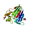
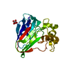
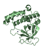
| Entry | Database: PDB / ID: 3iuy | ||||||
|---|---|---|---|---|---|---|---|
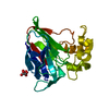
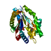
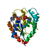
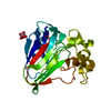
| Title | Crystal structure of DDX53 DEAD-box domain | ||||||
 Components Components | Probable ATP-dependent RNA helicase DDX53 | ||||||
 Keywords Keywords |  HYDROLASE / REC-A-like / DEAD-box / Structural Genomics / Structural Genomics Consortium / SGC / ATP-binding / Helicase / Nucleotide-binding / Nucleus / RNA-binding HYDROLASE / REC-A-like / DEAD-box / Structural Genomics / Structural Genomics Consortium / SGC / ATP-binding / Helicase / Nucleotide-binding / Nucleus / RNA-binding | ||||||
| Function / homology |  Function and homology informationRNA helicase activity / RNA helicase / intracellular membrane-bounded organelle / nucleolus / ATP hydrolysis activity / RNA binding / nucleoplasm / ATP binding / cytosol Function and homology informationRNA helicase activity / RNA helicase / intracellular membrane-bounded organelle / nucleolus / ATP hydrolysis activity / RNA binding / nucleoplasm / ATP binding / cytosolSimilarity search - Function | ||||||
| Biological species |  Homo sapiens (human) Homo sapiens (human) | ||||||
| Method | X-RAY DIFFRACTION / SYNCHROTRON / MOLECULAR REPLACEMENT / Resolution: 2.4 Å | ||||||
 Authors Authors | Schutz, P. / Karlberg, T. / Collins, R. / Arrowsmith, C.H. / Berglund, H. / Bountra, C. / Edwards, A.M. / Flodin, S. / Flores, A. / Graslund, S. ...Schutz, P. / Karlberg, T. / Collins, R. / Arrowsmith, C.H. / Berglund, H. / Bountra, C. / Edwards, A.M. / Flodin, S. / Flores, A. / Graslund, S. / Hammarstrom, M. / Johansson, A. / Johansson, I. / Kallas, A. / Kraulis, P. / Kotenyova, T. / Kotzsch, A. / Markova, N. / Moche, M. / Nielsen, T.K. / Nordlund, P. / Nyman, T. / Persson, C. / Roos, A.K. / Siponen, M.I. / Svensson, L. / Thorsell, A.G. / Tresaugues, L. / Van Den Berg, S. / Wahlberg, E. / Weigelt, J. / Welin, M. / Wisniewska, M. / Schuler, H.M. / Structural Genomics Consortium (SGC) | ||||||
 Citation Citation | Journal: Plos One / Year: 2010 Title: Comparative Structural Analysis of Human DEAD-Box RNA Helicases. Authors: Schutz, P. / Karlberg, T. / van den Berg, S. / Collins, R. / Lehtio, L. / Hogbom, M. / Holmberg-Schiavone, L. / Tempel, W. / Park, H.W. / Hammarstrom, M. / Moche, M. / Thorsell, A.G. / Schuler, H. | ||||||
| History |
|
- Structure visualization
| Structure viewer | Molecule: MolmilJmol/JSmol |
|---|
- Downloads & links
-Download
| PDBx/mmCIF format | 3iuy.cif.gz | 97.8 KB | Display | PDBx/mmCIF format |
|---|---|---|---|---|
| PDB format | pdb3iuy.ent.gz | 73.4 KB | Display | PDB format |
| PDBx/mmJSON format | 3iuy.json.gz | Tree view | PDBx/mmJSON format | |
| Others |  Other downloads Other downloads |
-Validation report
| Arichive directory | https://data.pdbj.org/pub/pdb/validation_reports/iu/3iuyftp://data.pdbj.org/pub/pdb/validation_reports/iu/3iuy | HTTPS FTP |
|---|
-Related structure data
| Related structure data | 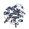 2g9nC 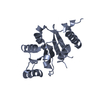 2p6nC  2pl3C  2rb4C 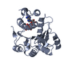 3b7gC 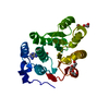 3berC  3borC 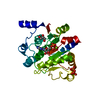 3dkpC 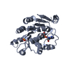 3fe2C  3ly5C C: citing same article ( |
|---|---|
| Similar structure data |


-Links
 PDBj
PDBj


- Assembly
| Deposited unit | 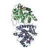
| ||||||||
|---|---|---|---|---|---|---|---|---|---|
| 1 | 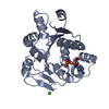
| ||||||||
| 2 | 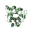
| ||||||||
| Unit cell |
|
-Components
| #1: Protein | Mass: 25750.002 Da / Num. of mol.: 2 / Fragment: UNP residues 204-430, Helicase ATP-binding domain Source method: isolated from a genetically manipulated source Source: (gene. exp.) Homo sapiens (human) / Gene: DDX53, CAGE / Plasmid: pNIC-Bsa4 / Production host:  Escherichia coli (E. coli) / Strain (production host): BL21(DE3) R3 pRARE Escherichia coli (E. coli) / Strain (production host): BL21(DE3) R3 pRAREReferences: UniProt: Q86TM3, Hydrolases; Acting on acid anhydrides; In phosphorus-containing anhydrides#2: Chemical | Adenosine monophosphate  Mass: 347.221 Da / Num. of mol.: 2 / Source method: obtained synthetically / Formula: C10H14N5O7P / Comment: AMP*YM Mass: 347.221 Da / Num. of mol.: 2 / Source method: obtained synthetically / Formula: C10H14N5O7P / Comment: AMP*YM#3: Chemical | Chloride  Mass: 35.453 Da / Num. of mol.: 2 / Source method: obtained synthetically / Formula: Cl Mass: 35.453 Da / Num. of mol.: 2 / Source method: obtained synthetically / Formula: Cl#4: Water | ChemComp-HOH / | Water Mass: 18.015 Da / Num. of mol.: 155 / Source method: isolated from a natural source / Formula: H2O Mass: 18.015 Da / Num. of mol.: 155 / Source method: isolated from a natural source / Formula: H2O |
|---|
-Experimental details
-Experiment
| Experiment | Method: X-RAY DIFFRACTION / Number of used crystals: 2 |
|---|
- Sample preparation
| Crystal | Density Matthews: 2.19 Å3/Da / Density % sol: 43.95 % |
|---|---|
| Crystal grow | Temperature: 277 K / Method: vapor diffusion, sitting drop / pH: 6 Details: 27% PEG6000, 0.1M MES pH 6, 0.2M AmmoniumChloride, 15% Glycerol, 0.2M NaCl , VAPOR DIFFUSION, SITTING DROP, temperature 277K |
-Data collection
| Diffraction source | Source: SYNCHROTRON / Site: ESRF  / Beamline: ID14-2 / Beamline: ID14-2 | ||||||||||||||||||||||||||||||||||||||||||||||||||||||||||||||||||||||||||||||||||||||||
|---|---|---|---|---|---|---|---|---|---|---|---|---|---|---|---|---|---|---|---|---|---|---|---|---|---|---|---|---|---|---|---|---|---|---|---|---|---|---|---|---|---|---|---|---|---|---|---|---|---|---|---|---|---|---|---|---|---|---|---|---|---|---|---|---|---|---|---|---|---|---|---|---|---|---|---|---|---|---|---|---|---|---|---|---|---|---|---|---|---|
| Radiation | Protocol: SINGLE WAVELENGTH / Monochromatic (M) / Laue (L): M / Scattering type: x-ray | ||||||||||||||||||||||||||||||||||||||||||||||||||||||||||||||||||||||||||||||||||||||||
| Radiation wavelength | Relative weight: 1 | ||||||||||||||||||||||||||||||||||||||||||||||||||||||||||||||||||||||||||||||||||||||||
| Reflection | Resolution: 2.4→65.382 Å / Num. obs: 17649 / % possible obs: 100 % / Redundancy: 9.3 % / Rmerge(I) obs: 0.187 / Rsym value: 0.187 / Net I/σ(I): 13.4 | ||||||||||||||||||||||||||||||||||||||||||||||||||||||||||||||||||||||||||||||||||||||||
| Reflection shell |
|
- Processing
| Software |
| |||||||||||||||||||||||||||||||||||||||||||||||||||||||||||||||||
|---|---|---|---|---|---|---|---|---|---|---|---|---|---|---|---|---|---|---|---|---|---|---|---|---|---|---|---|---|---|---|---|---|---|---|---|---|---|---|---|---|---|---|---|---|---|---|---|---|---|---|---|---|---|---|---|---|---|---|---|---|---|---|---|---|---|---|
| Refinement | Method to determine structure: MOLECULAR REPLACEMENT / Resolution: 2.4→45.13 Å / Cor.coef. Fo:Fc: 0.927 / Cor.coef. Fo:Fc free: 0.882 / Occupancy max: 1 / Occupancy min: 0 / SU B: 8.703 / SU ML: 0.203 / Cross valid method: THROUGHOUT / σ(F): 0 / ESU R: 0.515 / ESU R Free: 0.276 / Stereochemistry target values: MAXIMUM LIKELIHOOD Details: HYDROGENS HAVE BEEN ADDED IN THE RIDING POSITIONS U VALUES : REFINED INDIVIDUALLY
| |||||||||||||||||||||||||||||||||||||||||||||||||||||||||||||||||
| Solvent computation | Ion probe radii: 0.8 Å / Shrinkage radii: 0.8 Å / VDW probe radii: 1.2 Å / Solvent model: MASK | |||||||||||||||||||||||||||||||||||||||||||||||||||||||||||||||||
| Displacement parameters | Biso max: 56.43 Å2 / Biso mean: 23.168 Å2 / Biso min: 4.49 Å2
| |||||||||||||||||||||||||||||||||||||||||||||||||||||||||||||||||
| Refinement step | Cycle: LAST / Resolution: 2.4→45.13 Å
| |||||||||||||||||||||||||||||||||||||||||||||||||||||||||||||||||
| Refine LS restraints |
| |||||||||||||||||||||||||||||||||||||||||||||||||||||||||||||||||
| LS refinement shell | Resolution: 2.4→2.462 Å / Total num. of bins used: 20
|
