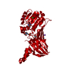
 Movie
Movie Controller

[English] 日本語
 Yorodumi
- PDB-2zqb: Crystal structure of a psychrotrophic RNaseHI variant with sextup... -
+ Open data

- Basic information
| Entry | Database: PDB / ID: 2zqb | ||||||
|---|---|---|---|---|---|---|---|
| Title | Crystal structure of a psychrotrophic RNaseHI variant with sextuple thermostabilizing mutations | ||||||
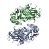
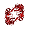
 Components Components | Ribonuclease HI | ||||||
 Keywords Keywords |  HYDROLASE / Cytoplasm / Endonuclease / Magnesium / Metal-binding / Nuclease HYDROLASE / Cytoplasm / Endonuclease / Magnesium / Metal-binding / Nuclease | ||||||
| Function / homology |  Function and homology informationDNA replication, removal of RNA primer / ribonuclease H / RNA-DNA hybrid ribonuclease activity / nucleic acid binding / magnesium ion binding / cytoplasm Function and homology informationDNA replication, removal of RNA primer / ribonuclease H / RNA-DNA hybrid ribonuclease activity / nucleic acid binding / magnesium ion binding / cytoplasmSimilarity search - Function | ||||||
| Biological species |  Shewanella oneidensis (bacteria) Shewanella oneidensis (bacteria) | ||||||
| Method | X-RAY DIFFRACTION / SYNCHROTRON / MOLECULAR REPLACEMENT / Resolution: 2.49 Å | ||||||
 Authors Authors | Angkawidjaja, C. / Kanaya, S. | ||||||
 Citation Citation | Journal: Febs J. / Year: 2009 Title: Destabilization of psychrotrophic RNase HI in a localized fashion as revealed by mutational and X-ray crystallographic analyses Authors: Rohman, M.S. / Tadokoro, T. / Angkawidjaja, C. / Abe, Y. / Matsumura, H. / Koga, Y. / Takano, K. / Kanaya, S. | ||||||
| History |
|
- Structure visualization
| Structure viewer | Molecule: MolmilJmol/JSmol |
|---|
- Downloads & links
-Download
| PDBx/mmCIF format | 2zqb.cif.gz | 130.6 KB | Display | PDBx/mmCIF format |
|---|---|---|---|---|
| PDB format | pdb2zqb.ent.gz | 103.3 KB | Display | PDB format |
| PDBx/mmJSON format | 2zqb.json.gz | Tree view | PDBx/mmJSON format | |
| Others |  Other downloads Other downloads |
-Validation report
| Arichive directory | https://data.pdbj.org/pub/pdb/validation_reports/zq/2zqbftp://data.pdbj.org/pub/pdb/validation_reports/zq/2zqb | HTTPS FTP |
|---|
-Related structure data
| Related structure data |  2e4lS S: Starting model for refinement |
|---|---|
| Similar structure data |


-Links
 PDBj
PDBj
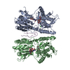
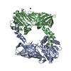
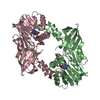
- Assembly
| Deposited unit | 
| ||||||||
|---|---|---|---|---|---|---|---|---|---|
| 1 |
| ||||||||
| Unit cell |
|
-Components
| #1: Protein | Mass: 17640.176 Da / Num. of mol.: 4 / Mutation: N29K, D39G, M76V, K90N, R97G, D136H Source method: isolated from a genetically manipulated source Source: (gene. exp.) Shewanella oneidensis (bacteria) / Strain: MR-1 / Plasmid: pET25b(+) / Production host: Escherichia coli (E. coli) / Strain (production host): MIC2067(DE3) / References: UniProt: Q8EE30, ribonuclease H#2: Chemical | Sulfate  Mass: 96.063 Da / Num. of mol.: 3 / Source method: obtained synthetically / Formula: SO4 Mass: 96.063 Da / Num. of mol.: 3 / Source method: obtained synthetically / Formula: SO4#3: Water | ChemComp-HOH / | Water Mass: 18.015 Da / Num. of mol.: 224 / Source method: isolated from a natural source / Formula: H2O Mass: 18.015 Da / Num. of mol.: 224 / Source method: isolated from a natural source / Formula: H2O |
|---|
-Experimental details
-Experiment
| Experiment | Method: X-RAY DIFFRACTION / Number of used crystals: 1 |
|---|
- Sample preparation
| Crystal | Density Matthews: 2.25 Å3/Da / Density % sol: 45.35 % |
|---|---|
| Crystal grow | Temperature: 277 K / Method: vapor diffusion, sitting drop / pH: 6.5 Details: 30% PEG, MME 5000, 0.1M MES, 0.2M ammonium sulfate, pH 6.5, VAPOR DIFFUSION, SITTING DROP, temperature 277K |
-Data collection
| Diffraction | Mean temperature: 100 K |
|---|---|
| Diffraction source | Source: SYNCHROTRON / Site: SPring-8  / Beamline: BL44XU / Wavelength: 1 Å / Beamline: BL44XU / Wavelength: 1 Å |
| Detector | Type: Bruker DIP-6040 / Detector: CCD / Date: Jul 2, 2008 / Details: mirrors |
| Radiation | Monochromator: mirror / Protocol: SINGLE WAVELENGTH / Monochromatic (M) / Laue (L): M / Scattering type: x-ray |
| Radiation wavelength | Wavelength: 1 Å / Relative weight: 1 |
| Reflection | Resolution: 2.49→50 Å / Num. obs: 23782 / % possible obs: 100 % / Redundancy: 13.9 % / Rmerge(I) obs: 0.146 / Net I/σ(I): 28.4 |
| Reflection shell | Resolution: 2.49→2.59 Å / Redundancy: 14.2 % / Rmerge(I) obs: 0.525 / Mean I/σ(I) obs: 6.39 / Num. unique all: 2299 / % possible all: 100 |
- Processing
| Software |
| ||||||||||||||||||||||||||||||||||||||||||||||||||||||||||||||||||||||||||||||||||||||||||
|---|---|---|---|---|---|---|---|---|---|---|---|---|---|---|---|---|---|---|---|---|---|---|---|---|---|---|---|---|---|---|---|---|---|---|---|---|---|---|---|---|---|---|---|---|---|---|---|---|---|---|---|---|---|---|---|---|---|---|---|---|---|---|---|---|---|---|---|---|---|---|---|---|---|---|---|---|---|---|---|---|---|---|---|---|---|---|---|---|---|---|---|
| Refinement | Method to determine structure: MOLECULAR REPLACEMENT Starting model: PDB ENTRY 2E4L Resolution: 2.49→47.51 Å / Cor.coef. Fo:Fc: 0.932 / Cor.coef. Fo:Fc free: 0.894 / SU B: 8.093 / SU ML: 0.186 / Cross valid method: THROUGHOUT / ESU R: 0.684 / ESU R Free: 0.295 Stereochemistry target values: MAXIMUM LIKELIHOOD WITH PHASES
| ||||||||||||||||||||||||||||||||||||||||||||||||||||||||||||||||||||||||||||||||||||||||||
| Solvent computation | Ion probe radii: 0.8 Å / Shrinkage radii: 0.8 Å / VDW probe radii: 1.2 Å / Solvent model: MASK | ||||||||||||||||||||||||||||||||||||||||||||||||||||||||||||||||||||||||||||||||||||||||||
| Displacement parameters | Biso mean: 28.584 Å2
| ||||||||||||||||||||||||||||||||||||||||||||||||||||||||||||||||||||||||||||||||||||||||||
| Refinement step | Cycle: LAST / Resolution: 2.49→47.51 Å
| ||||||||||||||||||||||||||||||||||||||||||||||||||||||||||||||||||||||||||||||||||||||||||
| Refine LS restraints |
| ||||||||||||||||||||||||||||||||||||||||||||||||||||||||||||||||||||||||||||||||||||||||||
| LS refinement shell | Resolution: 2.487→2.552 Å / Total num. of bins used: 20
|
