 Movie
Movie Controller

[English] 日本語
 Yorodumi
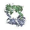
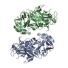
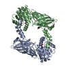
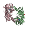
- PDB-5fub: Crystal Structure of zebrafish Protein Arginine Methyltransferase... -
+ Open data

- Basic information
| Entry | Database: PDB / ID: 5fub | ||||||
|---|---|---|---|---|---|---|---|
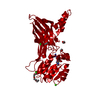
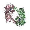
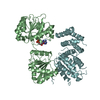
| Title | Crystal Structure of zebrafish Protein Arginine Methyltransferase 2 catalytic domain with SAH | ||||||
 Components Components | PROTEIN ARGININE METHYLTRANSFERASE 2 | ||||||
 Keywords Keywords |  TRANSFERASE / S-ADENOSYL-L-CYSTEINE / S-ADENOSYL-L-HOMOCYSTEINE TRANSFERASE / S-ADENOSYL-L-CYSTEINE / S-ADENOSYL-L-HOMOCYSTEINE | ||||||
| Function / homology |  Function and homology information Function and homology informationpeptidyl-arginine methylation / protein-arginine N-methyltransferase activity / S-adenosylmethionine-dependent methyltransferase activity / identical protein binding / nucleusSimilarity search - Function | ||||||
| Biological species |  DANIO RERIO (zebrafish) DANIO RERIO (zebrafish) | ||||||
| Method | X-RAY DIFFRACTION / SYNCHROTRON / MOLECULAR REPLACEMENT / Resolution: 1.997 Å | ||||||
 Authors Authors | Cura, V. / Troffer-Charlier, N. / Marechal, N. / Bonnefond, L. / Cavarelli, J. | ||||||
 Citation Citation | Journal: FEBS J. / Year: 2017 Title: Structural studies of protein arginine methyltransferase 2 reveal its interactions with potential substrates and inhibitors. Authors: Cura, V. / Marechal, N. / Troffer-Charlier, N. / Strub, J.M. / van Haren, M.J. / Martin, N.I. / Cianferani, S. / Bonnefond, L. / Cavarelli, J. | ||||||
| History |
|
- Structure visualization
| Structure viewer | Molecule: MolmilJmol/JSmol |
|---|
- Downloads & links
-Download
| PDBx/mmCIF format | 5fub.cif.gz | 148.1 KB | Display | PDBx/mmCIF format |
|---|---|---|---|---|
| PDB format | pdb5fub.ent.gz | 116.5 KB | Display | PDB format |
| PDBx/mmJSON format | 5fub.json.gz | Tree view | PDBx/mmJSON format | |
| Others |  Other downloads Other downloads |
-Validation report
| Arichive directory | https://data.pdbj.org/pub/pdb/validation_reports/fu/5fubftp://data.pdbj.org/pub/pdb/validation_reports/fu/5fub | HTTPS FTP |
|---|
-Related structure data
| Related structure data | 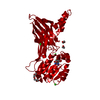 5fulC  5fwaC 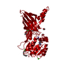 5fwdC 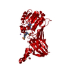 5g02C 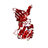 5jmqC 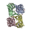 5k8vC C: citing same article ( |
|---|---|
| Similar structure data |


-Links
 PDBj
PDBj- Assembly
| Deposited unit | 
| ||||||||
|---|---|---|---|---|---|---|---|---|---|
| 1 | 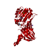
| ||||||||
| Unit cell |
|
-Components
-Protein , 1 types, 1 molecules A
| #1: Protein | Mass: 38376.770 Da / Num. of mol.: 1 / Fragment: CATALYTIC MODULE, UNP RESIDUES 73-408 Source method: isolated from a genetically manipulated source Source: (gene. exp.) DANIO RERIO (zebrafish) / Description: BIOSCIENCE IRAK293-C16 / Plasmid: PDEST20 / Cell line (production host): Sf21 / Production host:   SPODOPTERA FRUGIPERDA (fall armyworm) / References: UniProt: A1L1Q4, EC: 2.1.1.125 SPODOPTERA FRUGIPERDA (fall armyworm) / References: UniProt: A1L1Q4, EC: 2.1.1.125 |
|---|
-Non-polymers , 6 types, 190 molecules 


| #2: Chemical | ChemComp-SAH / S-Adenosyl-L-homocysteine Type: L-peptide linking / Mass: 384.411 Da / Num. of mol.: 1 / Source method: obtained synthetically / Formula: C14H20N6O5S Type: L-peptide linking / Mass: 384.411 Da / Num. of mol.: 1 / Source method: obtained synthetically / Formula: C14H20N6O5S | ||||
|---|---|---|---|---|---|
| #3: Chemical | ChemComp-MES / MES (buffer) Mass: 195.237 Da / Num. of mol.: 1 / Source method: obtained synthetically / Formula: C6H13NO4S / Comment: pH buffer*YM Mass: 195.237 Da / Num. of mol.: 1 / Source method: obtained synthetically / Formula: C6H13NO4S / Comment: pH buffer*YM | ||||
| #4: Chemical | ChemComp-CL / Chloride Mass: 35.453 Da / Num. of mol.: 1 / Source method: obtained synthetically / Formula: Cl Mass: 35.453 Da / Num. of mol.: 1 / Source method: obtained synthetically / Formula: Cl | ||||
| #5: Chemical |  Mass: 22.990 Da / Num. of mol.: 2 / Source method: obtained synthetically / Formula: Na Mass: 22.990 Da / Num. of mol.: 2 / Source method: obtained synthetically / Formula: Na#6: Chemical | ChemComp-EDO / Ethylene glycol Mass: 62.068 Da / Num. of mol.: 4 / Source method: obtained synthetically / Formula: C2H6O2 Mass: 62.068 Da / Num. of mol.: 4 / Source method: obtained synthetically / Formula: C2H6O2#7: Water | ChemComp-HOH / | WaterMass: 18.015 Da / Num. of mol.: 181 / Source method: isolated from a natural source / Formula: H2O |
-Details
| Sequence details | CRYSTALLIZ |
|---|
-Experimental details
-Experiment
| Experiment | Method: X-RAY DIFFRACTION / Number of used crystals: 1 |
|---|
- Sample preparation
| Crystal | Density Matthews: 3.42 Å3/Da / Density % sol: 64 % / Description: STARTING MODEL GENERATED BY BALBES |
|---|---|
| Crystal grow | pH: 6 Details: 9% PEG 20000, 300MM NACL,100MM MES PH 6.0 AND FOR CRYO IS 10% PEG 20000, 300MM NACL,100MM MES PH 6.0, 15% ETHYLENE GLYCOL |
-Data collection
| Diffraction | Mean temperature: 100 K |
|---|---|
| Diffraction source | Source: SYNCHROTRON / Site: SOLEIL  / Beamline: PROXIMA 2 / Wavelength: 0.9801 / Beamline: PROXIMA 2 / Wavelength: 0.9801 |
| Detector | Type: ADSC QUANTUM 315 / Detector: CCD / Date: Apr 3, 2014 |
| Radiation | Protocol: SINGLE WAVELENGTH / Monochromatic (M) / Laue (L): M / Scattering type: x-ray |
| Radiation wavelength | Wavelength: 0.9801 Å / Relative weight: 1 |
| Reflection | Resolution: 2→38.4 Å / Num. obs: 35917 / % possible obs: 99.9 % / Observed criterion σ(I): 0 / Redundancy: 5.3 % / Biso Wilson estimate: 35.3 Å2 / Rmerge(I) obs: 0.1 / Net I/σ(I): 9.2 |
| Reflection shell | Resolution: 2→2.05 Å / Redundancy: 5.3 % / Mean I/σ(I) obs: 1 / % possible all: 100 |
- Processing
| Software |
| ||||||||||||||||||||||||||||||||||||||||||||||||||||||||||||||||||||||||||||||||||||||||||||||||||||||||||||||||||||||||||||||||||||||||||||||||||||||
|---|---|---|---|---|---|---|---|---|---|---|---|---|---|---|---|---|---|---|---|---|---|---|---|---|---|---|---|---|---|---|---|---|---|---|---|---|---|---|---|---|---|---|---|---|---|---|---|---|---|---|---|---|---|---|---|---|---|---|---|---|---|---|---|---|---|---|---|---|---|---|---|---|---|---|---|---|---|---|---|---|---|---|---|---|---|---|---|---|---|---|---|---|---|---|---|---|---|---|---|---|---|---|---|---|---|---|---|---|---|---|---|---|---|---|---|---|---|---|---|---|---|---|---|---|---|---|---|---|---|---|---|---|---|---|---|---|---|---|---|---|---|---|---|---|---|---|---|---|---|---|---|
| Refinement | Method to determine structure: MOLECULAR REPLACEMENT Starting model: NONE Resolution: 1.997→38.405 Å / SU ML: 0.25 / σ(F): 1.36 / Phase error: 22.6 / Stereochemistry target values: ML
| ||||||||||||||||||||||||||||||||||||||||||||||||||||||||||||||||||||||||||||||||||||||||||||||||||||||||||||||||||||||||||||||||||||||||||||||||||||||
| Solvent computation | Shrinkage radii: 0.9 Å / VDW probe radii: 1.11 Å / Solvent model: FLAT BULK SOLVENT MODEL | ||||||||||||||||||||||||||||||||||||||||||||||||||||||||||||||||||||||||||||||||||||||||||||||||||||||||||||||||||||||||||||||||||||||||||||||||||||||
| Displacement parameters | Biso mean: 45.6 Å2 | ||||||||||||||||||||||||||||||||||||||||||||||||||||||||||||||||||||||||||||||||||||||||||||||||||||||||||||||||||||||||||||||||||||||||||||||||||||||
| Refinement step | Cycle: LAST / Resolution: 1.997→38.405 Å
| ||||||||||||||||||||||||||||||||||||||||||||||||||||||||||||||||||||||||||||||||||||||||||||||||||||||||||||||||||||||||||||||||||||||||||||||||||||||
| Refine LS restraints |
| ||||||||||||||||||||||||||||||||||||||||||||||||||||||||||||||||||||||||||||||||||||||||||||||||||||||||||||||||||||||||||||||||||||||||||||||||||||||
| LS refinement shell |
| ||||||||||||||||||||||||||||||||||||||||||||||||||||||||||||||||||||||||||||||||||||||||||||||||||||||||||||||||||||||||||||||||||||||||||||||||||||||
| Refinement TLS params. | Method: refined / Refine-ID: X-RAY DIFFRACTION
| ||||||||||||||||||||||||||||||||||||||||||||||||||||||||||||||||||||||||||||||||||||||||||||||||||||||||||||||||||||||||||||||||||||||||||||||||||||||
| Refinement TLS group |
|
