 Movie
Movie Controller

[English] 日本語
 Yorodumi
- PDB-2z48: Crystal Structure of Hemolytic Lectin CEL-III Complexed with GalNac -
+ Open data

- Basic information
| Entry | Database: PDB / ID: 2z48 | ||||||||||||
|---|---|---|---|---|---|---|---|---|---|---|---|---|---|
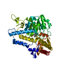
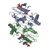
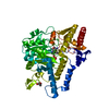
| Title | Crystal Structure of Hemolytic Lectin CEL-III Complexed with GalNac | ||||||||||||
 Components Components | Hemolytic lectin CEL-III | ||||||||||||
 Keywords Keywords |  TOXIN / Lectin / CEL-III / Hemolysis / Hemagglutination / Pore-forming / Calcium / Magnesium / GalNac TOXIN / Lectin / CEL-III / Hemolysis / Hemagglutination / Pore-forming / Calcium / Magnesium / GalNac | ||||||||||||
| Function / homology |  Function and homology information Function and homology informationpositive regulation of erythrocyte aggregation / melibiose binding / lactose binding / cell killing / N-acetylgalactosamine binding / fucose binding / disruption of plasma membrane integrity in another organism / galactose binding / hemolysis in another organism / protein homooligomerization ...positive regulation of erythrocyte aggregation / melibiose binding / lactose binding / cell killing / N-acetylgalactosamine binding / fucose binding / disruption of plasma membrane integrity in another organism / galactose binding / hemolysis in another organism / protein homooligomerization / : / antibacterial humoral response / defense response to Gram-negative bacterium / defense response to Gram-positive bacterium / calcium ion binding / magnesium ion binding / extracellular spaceSimilarity search - Function | ||||||||||||
| Biological species |  Cucumaria echinata (invertebrata) Cucumaria echinata (invertebrata) | ||||||||||||
| Method | X-RAY DIFFRACTION / SYNCHROTRON / MOLECULAR REPLACEMENT / Resolution: 1.7 Å | ||||||||||||
 Authors Authors | Hatakeyama, T. / Unno, H. / Eto, S. / Hidemura, H. / Uchida, T. / Kouzuma, Y. | ||||||||||||
 Citation Citation | Journal: J.Biol.Chem. / Year: 2007 Title: C-type lectin-like carbohydrate-recognition of the hemolytic lectin CEL-III containing ricin-type beta-trefoil folds Authors: Hatakeyama, T. / Unno, H. / Kouzuma, Y. / Uchida, T. / Eto, S. / Hidemura, H. / Kato, N. / Yonekura, M. / Kusunoki, M. | ||||||||||||
| History |
|
- Structure visualization
| Structure viewer | Molecule: MolmilJmol/JSmol |
|---|
- Downloads & links
-Download
| PDBx/mmCIF format | 2z48.cif.gz | 203.2 KB | Display | PDBx/mmCIF format |
|---|---|---|---|---|
| PDB format | pdb2z48.ent.gz | 158.6 KB | Display | PDB format |
| PDBx/mmJSON format | 2z48.json.gz | Tree view | PDBx/mmJSON format | |
| Others |  Other downloads Other downloads |
-Validation report
| Arichive directory | https://data.pdbj.org/pub/pdb/validation_reports/z4/2z48ftp://data.pdbj.org/pub/pdb/validation_reports/z4/2z48 | HTTPS FTP |
|---|
-Related structure data
| Related structure data |  2z49C  1vclS S: Starting model for refinement C: citing same article ( |
|---|---|
| Similar structure data |


-Links
 PDBj
PDBj

- Assembly
| Deposited unit | 
| ||||||||||||||||||
|---|---|---|---|---|---|---|---|---|---|---|---|---|---|---|---|---|---|---|---|
| 1 | 
| ||||||||||||||||||
| 2 | 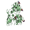
| ||||||||||||||||||
| Unit cell |
| ||||||||||||||||||
| Noncrystallographic symmetry (NCS) | NCS domain:
NCS domain segments: Component-ID: 1 / Ens-ID: 1 / Beg auth comp-ID: VAL / Beg label comp-ID: VAL / End auth comp-ID: ILE / End label comp-ID: ILE / Refine code: 1 / Auth seq-ID: 2 - 432 / Label seq-ID: 2 - 432
|
-Components
-Protein , 1 types, 2 molecules AB
| #1: Protein | Mass: 47452.164 Da / Num. of mol.: 2 / Fragment: residues in database 11-442 / Mutation: Q11PCA Source method: isolated from a genetically manipulated source Source: (gene. exp.) Cucumaria echinata (invertebrata) / Production host: Escherichia coli / References: UniProt: Q868M7 |
|---|
-Sugars , 2 types, 12 molecules 
| #2: Sugar | ChemComp-A2G / N-Acetylgalactosamine Type: D-saccharide, alpha linking / Mass: 221.208 Da / Num. of mol.: 4 Type: D-saccharide, alpha linking / Mass: 221.208 Da / Num. of mol.: 4Source method: isolated from a genetically manipulated source Formula: C8H15NO6 #3: Sugar | ChemComp-NGA / N-Acetylgalactosamine Type: D-saccharide, beta linking / Mass: 221.208 Da / Num. of mol.: 8 Type: D-saccharide, beta linking / Mass: 221.208 Da / Num. of mol.: 8Source method: isolated from a genetically manipulated source Formula: C8H15NO6 |
|---|
-Non-polymers , 3 types, 852 molecules 


| #4: Chemical | ChemComp-CA /  Mass: 40.078 Da / Num. of mol.: 10 / Source method: obtained synthetically / Formula: Ca Mass: 40.078 Da / Num. of mol.: 10 / Source method: obtained synthetically / Formula: Ca#5: Chemical | ChemComp-MG /  Mass: 24.305 Da / Num. of mol.: 4 / Source method: obtained synthetically / Formula: Mg Mass: 24.305 Da / Num. of mol.: 4 / Source method: obtained synthetically / Formula: Mg#6: Water | ChemComp-HOH / | WaterMass: 18.015 Da / Num. of mol.: 838 / Source method: isolated from a natural source / Formula: H2O |
|---|
-Details
| Sequence details | THERE ARE 13 DIFFERENCES BETWEEN THE DEPOSITORS DATA AND THE GENETIC SEQUENCE. THERE IS NO QUESTION ...THERE ARE 13 DIFFERENCE |
|---|
-Experimental details
-Experiment
| Experiment | Method: X-RAY DIFFRACTION / Number of used crystals: 1 |
|---|
- Sample preparation
| Crystal | Density Matthews: 2.3 Å3/Da / Density % sol: 46.58 % |
|---|---|
| Crystal grow | Temperature: 293 K / Method: vapor diffusion, sitting drop / pH: 6.5 Details: 10mM GalNac, 12% PEG 8000, 100mM Bis-Tris/NaOH, 200mM magnesium acetate, pH 6.5, VAPOR DIFFUSION, SITTING DROP, temperature 293K |
-Data collection
| Diffraction | Mean temperature: 100 K |
|---|---|
| Diffraction source | Source: SYNCHROTRON / Site: SPring-8  / Beamline: BL44XU / Wavelength: 0.9 Å / Beamline: BL44XU / Wavelength: 0.9 Å |
| Detector | Type: BRUKER DIP-6040B / Detector: IMAGE PLATE / Date: Oct 2, 2004 |
| Radiation | Protocol: SINGLE WAVELENGTH / Monochromatic (M) / Laue (L): M / Scattering type: x-ray |
| Radiation wavelength | Wavelength: 0.9 Å / Relative weight: 1 |
| Reflection | Resolution: 1.7→64.96 Å / Num. obs: 89844 / % possible obs: 96.4 % / Redundancy: 2 % / Biso Wilson estimate: 19.4 Å2 / Rmerge(I) obs: 0.067 / Net I/σ(I): 10.6 |
| Reflection shell | Resolution: 1.7→1.79 Å / Redundancy: 2 % / Rmerge(I) obs: 0.166 / Mean I/σ(I) obs: 3.5 / % possible all: 95.2 |
- Processing
| Software |
| ||||||||||||||||||||||||||||||||||||||||||||||||||||||||||||||||||||||||||||||||||||||||||||||||||||||||||||||||||||||||||||||||||||||||||||||||||||||||||||||||||||||||||
|---|---|---|---|---|---|---|---|---|---|---|---|---|---|---|---|---|---|---|---|---|---|---|---|---|---|---|---|---|---|---|---|---|---|---|---|---|---|---|---|---|---|---|---|---|---|---|---|---|---|---|---|---|---|---|---|---|---|---|---|---|---|---|---|---|---|---|---|---|---|---|---|---|---|---|---|---|---|---|---|---|---|---|---|---|---|---|---|---|---|---|---|---|---|---|---|---|---|---|---|---|---|---|---|---|---|---|---|---|---|---|---|---|---|---|---|---|---|---|---|---|---|---|---|---|---|---|---|---|---|---|---|---|---|---|---|---|---|---|---|---|---|---|---|---|---|---|---|---|---|---|---|---|---|---|---|---|---|---|---|---|---|---|---|---|---|---|---|---|---|---|---|
| Refinement | Method to determine structure: MOLECULAR REPLACEMENT Starting model: 1VCL Resolution: 1.7→64.96 Å / Cor.coef. Fo:Fc: 0.955 / Cor.coef. Fo:Fc free: 0.94 / SU B: 2.14 / SU ML: 0.072 / Cross valid method: THROUGHOUT / ESU R: 0.121 / ESU R Free: 0.114 / Stereochemistry target values: MAXIMUM LIKELIHOOD / Details: HYDROGENS HAVE BEEN ADDED IN THE RIDING POSITIONS
| ||||||||||||||||||||||||||||||||||||||||||||||||||||||||||||||||||||||||||||||||||||||||||||||||||||||||||||||||||||||||||||||||||||||||||||||||||||||||||||||||||||||||||
| Solvent computation | Ion probe radii: 0.8 Å / Shrinkage radii: 0.8 Å / VDW probe radii: 1.2 Å / Solvent model: MASK | ||||||||||||||||||||||||||||||||||||||||||||||||||||||||||||||||||||||||||||||||||||||||||||||||||||||||||||||||||||||||||||||||||||||||||||||||||||||||||||||||||||||||||
| Displacement parameters | Biso mean: 20.532 Å2
| ||||||||||||||||||||||||||||||||||||||||||||||||||||||||||||||||||||||||||||||||||||||||||||||||||||||||||||||||||||||||||||||||||||||||||||||||||||||||||||||||||||||||||
| Refinement step | Cycle: LAST / Resolution: 1.7→64.96 Å
| ||||||||||||||||||||||||||||||||||||||||||||||||||||||||||||||||||||||||||||||||||||||||||||||||||||||||||||||||||||||||||||||||||||||||||||||||||||||||||||||||||||||||||
| Refine LS restraints |
| ||||||||||||||||||||||||||||||||||||||||||||||||||||||||||||||||||||||||||||||||||||||||||||||||||||||||||||||||||||||||||||||||||||||||||||||||||||||||||||||||||||||||||
| Refine LS restraints NCS | Dom-ID: 1 / Auth asym-ID: A / Ens-ID: 1 / Number: 3306 / Refine-ID: X-RAY DIFFRACTION
| ||||||||||||||||||||||||||||||||||||||||||||||||||||||||||||||||||||||||||||||||||||||||||||||||||||||||||||||||||||||||||||||||||||||||||||||||||||||||||||||||||||||||||
| LS refinement shell | Resolution: 1.7→1.744 Å / Total num. of bins used: 20
|
