| Entry | Database: PDB / ID: 1vcl
|
|---|
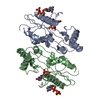
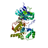
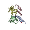
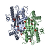
| Title | Crystal Structure of Hemolytic Lectin CEL-III |
|---|
 Components Components | hemolytic lectin CEL-III |
|---|
 Keywords Keywords |  TOXIN / LECTIN / CEL-III / HEMOLYSIS / HEMAGGLUTINATION / PORE-FORMING / CALCIUM TOXIN / LECTIN / CEL-III / HEMOLYSIS / HEMAGGLUTINATION / PORE-FORMING / CALCIUM |
|---|
| Function / homology |  Function and homology information Function and homology information
positive regulation of erythrocyte aggregation / melibiose binding / lactose binding / cell killing / N-acetylgalactosamine binding / fucose binding / disruption of plasma membrane integrity in another organism / galactose binding / hemolysis in another organism / protein homooligomerization ...positive regulation of erythrocyte aggregation / melibiose binding / lactose binding / cell killing / N-acetylgalactosamine binding / fucose binding / disruption of plasma membrane integrity in another organism / galactose binding / hemolysis in another organism / protein homooligomerization / : / antibacterial humoral response / defense response to Gram-negative bacterium / defense response to Gram-positive bacterium / calcium ion binding / magnesium ion binding / extracellular spaceSimilarity search - Function hemolytic lectin cel-iii, domain 3 / Hemolytic lectin CEL-III, C-terminal domain / Hemolytic lectin CEL-III, C-terminal domain superfamily / Ricin-type beta-trefoil lectin domain / Ricin-type beta-trefoil / Lectin domain of ricin B chain profile. / Ricin B, lectin domain / Ricin B-like lectins / Trefoil (Acidic Fibroblast Growth Factor, subunit A) - #50 / Trefoil (Acidic Fibroblast Growth Factor, subunit A) ...hemolytic lectin cel-iii, domain 3 / Hemolytic lectin CEL-III, C-terminal domain / Hemolytic lectin CEL-III, C-terminal domain superfamily / Ricin-type beta-trefoil lectin domain / Ricin-type beta-trefoil / Lectin domain of ricin B chain profile. / Ricin B, lectin domain / Ricin B-like lectins / Trefoil (Acidic Fibroblast Growth Factor, subunit A) - #50 / Trefoil (Acidic Fibroblast Growth Factor, subunit A) / Trefoil / 2-Layer Sandwich / Mainly Beta / Alpha BetaSimilarity search - Domain/homology |
|---|
| Biological species |  Cucumaria echinata (invertebrata) Cucumaria echinata (invertebrata) |
|---|
| Method | X-RAY DIFFRACTION / SYNCHROTRON / SIRAS / Resolution: 1.7 Å |
|---|
 Authors Authors | Uchida, T. / Yamasaki, T. / Eto, S. / Sugawara, H. / Kurisu, G. / Nakagawa, A. / Kusunoki, M. / Hatakeyama, T. |
|---|
 Citation Citation | Journal: J.Biol.Chem. / Year: 2004
Title: Crystal Structure of the Hemolytic Lectin CEL-III Isolated from the Marine Invertebrate Cucumaria echinata: IMPLICATIONS OF DOMAIN STRUCTURE FOR ITS MEMBRANE PORE-FORMATION MECHANISM
Authors: Uchida, T. / Yamasaki, T. / Eto, S. / Sugawara, H. / Kurisu, G. / Nakagawa, A. / Kusunoki, M. / Hatakeyama, T. |
|---|
| History | | Deposition | Mar 9, 2004 | Deposition site: PDBJ / Processing site: PDBJ |
|---|
| Revision 1.0 | Sep 7, 2004 | Provider: repository / Type: Initial release |
|---|
| Revision 1.1 | Apr 27, 2008 | Group: Version format compliance |
|---|
| Revision 1.2 | Jul 13, 2011 | Group: Version format compliance |
|---|
| Revision 2.0 | Dec 25, 2019 | Group: Database references / Derived calculations / Polymer sequence
Category: entity_poly / pdbx_struct_mod_residue ...entity_poly / pdbx_struct_mod_residue / struct_conn / struct_ref_seq_dif
Item: _entity_poly.pdbx_seq_one_letter_code_can / _pdbx_struct_mod_residue.parent_comp_id / _struct_conn.pdbx_leaving_atom_flag |
|---|
| Revision 2.1 | Dec 27, 2023 | Group: Data collection / Database references / Derived calculations
Category: chem_comp_atom / chem_comp_bond ...chem_comp_atom / chem_comp_bond / database_2 / pdbx_struct_conn_angle / struct_conn / struct_conn_type / struct_site
Item: _database_2.pdbx_DOI / _database_2.pdbx_database_accession ..._database_2.pdbx_DOI / _database_2.pdbx_database_accession / _pdbx_struct_conn_angle.ptnr1_auth_comp_id / _pdbx_struct_conn_angle.ptnr1_auth_seq_id / _pdbx_struct_conn_angle.ptnr1_label_asym_id / _pdbx_struct_conn_angle.ptnr1_label_atom_id / _pdbx_struct_conn_angle.ptnr1_label_comp_id / _pdbx_struct_conn_angle.ptnr1_label_seq_id / _pdbx_struct_conn_angle.ptnr2_auth_comp_id / _pdbx_struct_conn_angle.ptnr2_auth_seq_id / _pdbx_struct_conn_angle.ptnr2_label_asym_id / _pdbx_struct_conn_angle.ptnr2_label_atom_id / _pdbx_struct_conn_angle.ptnr2_label_comp_id / _pdbx_struct_conn_angle.ptnr3_auth_comp_id / _pdbx_struct_conn_angle.ptnr3_auth_seq_id / _pdbx_struct_conn_angle.ptnr3_label_asym_id / _pdbx_struct_conn_angle.ptnr3_label_atom_id / _pdbx_struct_conn_angle.ptnr3_label_comp_id / _pdbx_struct_conn_angle.ptnr3_label_seq_id / _pdbx_struct_conn_angle.value / _struct_conn.conn_type_id / _struct_conn.id / _struct_conn.pdbx_dist_value / _struct_conn.pdbx_leaving_atom_flag / _struct_conn.ptnr1_auth_asym_id / _struct_conn.ptnr1_auth_comp_id / _struct_conn.ptnr1_auth_seq_id / _struct_conn.ptnr1_label_asym_id / _struct_conn.ptnr1_label_atom_id / _struct_conn.ptnr1_label_comp_id / _struct_conn.ptnr1_label_seq_id / _struct_conn.ptnr2_auth_asym_id / _struct_conn.ptnr2_auth_comp_id / _struct_conn.ptnr2_auth_seq_id / _struct_conn.ptnr2_label_asym_id / _struct_conn.ptnr2_label_atom_id / _struct_conn.ptnr2_label_comp_id / _struct_conn.ptnr2_label_seq_id / _struct_conn_type.id / _struct_site.pdbx_auth_asym_id / _struct_site.pdbx_auth_comp_id / _struct_site.pdbx_auth_seq_id |
|---|
|
|---|
 Movie
Movie Controller


 Open data
 Basic information
 Structure visualization
 Downloads & links
 Other downloads
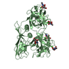


 PDBj
PDBj

 Assembly
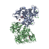


 Mass: 40.078 Da / Num. of mol.: 10 / Source method: obtained synthetically / Formula: Ca
 Mass: 24.305 Da / Num. of mol.: 4 / Source method: obtained synthetically / Formula: Mg
 Mass: 35.453 Da / Num. of mol.: 2 / Source method: obtained synthetically / Formula: Cl
 Mass: 209.240 Da / Num. of mol.: 1 / Source method: obtained synthetically / Formula: C8H19NO5 / Comment: pH buffer*YM
 Sample preparation
 / Beamline: BL-6A / Wavelength: 1 Å
 Processing