 Movie
Movie Controller

[English] 日本語
 Yorodumi
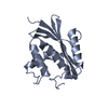
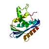
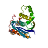
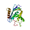
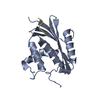
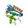
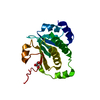
- PDB-2z1i: Crystal structure of E.coli RNase HI surface charged mutant(Q4R/T... -
+ Open data

- Basic information
| Entry | Database: PDB / ID: 2z1i | ||||||
|---|---|---|---|---|---|---|---|
| Title | Crystal structure of E.coli RNase HI surface charged mutant(Q4R/T40E/Q72H/Q76K/Q80E/T92K/Q105K/Q113R/Q115K) | ||||||
 Components Components | Ribonuclease HI | ||||||
 Keywords Keywords |  HYDROLASE / RNase HI / thermostability / surface-charge residue HYDROLASE / RNase HI / thermostability / surface-charge residue | ||||||
| Function / homology |  Function and homology informationDNA replication, removal of RNA primer / ribonuclease H / RNA-DNA hybrid ribonuclease activity / endonuclease activity / nucleic acid binding / magnesium ion binding / cytoplasm Function and homology informationDNA replication, removal of RNA primer / ribonuclease H / RNA-DNA hybrid ribonuclease activity / endonuclease activity / nucleic acid binding / magnesium ion binding / cytoplasmSimilarity search - Function | ||||||
| Biological species |  Escherichia coli (E. coli) Escherichia coli (E. coli) | ||||||
| Method | X-RAY DIFFRACTION / SYNCHROTRON / MOLECULAR REPLACEMENT / Resolution: 2 Å | ||||||
 Authors Authors | You, D.J. / Fukuchi, S. / Nishikawa, K. / Koga, Y. / Takano, K. / Kanaya, S. | ||||||
 Citation Citation | Journal: J.Biochem.(Tokyo) / Year: 2007 Title: Protein Thermostabilization Requires a Fine-tuned Placement of Surface-charged Residues Authors: You, D.-J. / Fukuchi, S. / Nishikawa, K. / Koga, Y. / Takano, K. / Kanaya, S. | ||||||
| History |
|
- Structure visualization
| Structure viewer | Molecule: MolmilJmol/JSmol |
|---|
- Downloads & links
-Download
| PDBx/mmCIF format | 2z1i.cif.gz | 78.5 KB | Display | PDBx/mmCIF format |
|---|---|---|---|---|
| PDB format | pdb2z1i.ent.gz | 58.4 KB | Display | PDB format |
| PDBx/mmJSON format | 2z1i.json.gz | Tree view | PDBx/mmJSON format | |
| Others |  Other downloads Other downloads |
-Validation report
| Arichive directory | https://data.pdbj.org/pub/pdb/validation_reports/z1/2z1iftp://data.pdbj.org/pub/pdb/validation_reports/z1/2z1i | HTTPS FTP |
|---|
-Related structure data
| Related structure data | 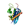 2z1gC 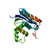 2z1hC 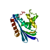 2z1jC 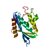 2rn2S C: citing same article ( S: Starting model for refinement |
|---|---|
| Similar structure data |


-Links
 PDBj
PDBj
- Assembly
| Deposited unit | 
| ||||||||
|---|---|---|---|---|---|---|---|---|---|
| 1 | 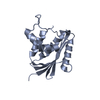
| ||||||||
| 2 | 
| ||||||||
| Unit cell |
|
-Components
| #1: Protein | Mass: 17751.375 Da / Num. of mol.: 2 / Mutation: Q4R/T40E/Q72H/Q76K/Q80E/T92K/Q105K/Q113R/Q115K Source method: isolated from a genetically manipulated source Source: (gene. exp.) Escherichia coli (E. coli) / Plasmid: pET25b / Production host: Escherichia coli (E. coli) / Strain (production host): MIC2067(DE3) / References: UniProt: P0A7Y4, ribonuclease H#2: Water | ChemComp-HOH / | Water Mass: 18.015 Da / Num. of mol.: 289 / Source method: isolated from a natural source / Formula: H2O Mass: 18.015 Da / Num. of mol.: 289 / Source method: isolated from a natural source / Formula: H2O |
|---|
-Experimental details
-Experiment
| Experiment | Method: X-RAY DIFFRACTION / Number of used crystals: 1 |
|---|
- Sample preparation
| Crystal | Density Matthews: 2.08 Å3/Da / Density % sol: 40.87 % |
|---|---|
| Crystal grow | Temperature: 293 K / Method: vapor diffusion, sitting drop / pH: 8.5 Details: 100mM HEPES-NaOH, 15-25% PEG 3350, pH 8.5, VAPOR DIFFUSION, SITTING DROP, temperature 293K |
-Data collection
| Diffraction | Mean temperature: 100 K |
|---|---|
| Diffraction source | Source: SYNCHROTRON / Site: SPring-8  / Beamline: BL44XU / Wavelength: 0.9 Å / Beamline: BL44XU / Wavelength: 0.9 Å |
| Detector | Type: BRUKER SMART 6500 / Detector: CCD / Date: Oct 3, 2005 |
| Radiation | Protocol: SINGLE WAVELENGTH / Monochromatic (M) / Laue (L): M / Scattering type: x-ray |
| Radiation wavelength | Wavelength: 0.9 Å / Relative weight: 1 |
| Reflection | Resolution: 2→50 Å / Num. obs: 20535 / % possible obs: 98.4 % / Biso Wilson estimate: 13.3 Å2 / Rmerge(I) obs: 0.098 / Net I/σ(I): 21.5 |
| Reflection shell | Resolution: 2→2.07 Å / Rmerge(I) obs: 0.231 / Mean I/σ(I) obs: 4.9 / % possible all: 91.4 |
- Processing
| Software |
| ||||||||||||||||||||||||||||||||||||
|---|---|---|---|---|---|---|---|---|---|---|---|---|---|---|---|---|---|---|---|---|---|---|---|---|---|---|---|---|---|---|---|---|---|---|---|---|---|
| Refinement | Method to determine structure: MOLECULAR REPLACEMENT Starting model: PDB ENTRY 2RN2 Resolution: 2→36.09 Å / Rfactor Rfree error: 0.008 / Data cutoff high absF: 1152282.49 / Data cutoff low absF: 0 / Isotropic thermal model: RESTRAINED / Cross valid method: THROUGHOUT / σ(F): 0
| ||||||||||||||||||||||||||||||||||||
| Solvent computation | Solvent model: FLAT MODEL / Bsol: 53.9535 Å2 / ksol: 0.352701 e/Å3 | ||||||||||||||||||||||||||||||||||||
| Displacement parameters | Biso mean: 30.7 Å2
| ||||||||||||||||||||||||||||||||||||
| Refine analyze |
| ||||||||||||||||||||||||||||||||||||
| Refinement step | Cycle: LAST / Resolution: 2→36.09 Å
| ||||||||||||||||||||||||||||||||||||
| Refine LS restraints |
| ||||||||||||||||||||||||||||||||||||
| LS refinement shell | Resolution: 2→2.13 Å / Rfactor Rfree error: 0.023 / Total num. of bins used: 6
| ||||||||||||||||||||||||||||||||||||
| Xplor file |
|
