 Movie
Movie Controller


+ Open data

- Basic information
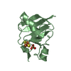
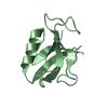
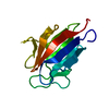
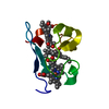
| Entry | Database: PDB / ID: 2ygs | ||||||
|---|---|---|---|---|---|---|---|
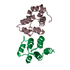
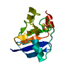
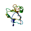
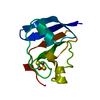
| Title | CARD DOMAIN FROM APAF-1 | ||||||
 Components Components | APOPTOTIC PROTEASE ACTIVATING FACTOR 1 | ||||||
 Keywords Keywords |  APOPTOSIS / CASPASE RECRUITMENT DOMAIN / APAF-1 / RECOGNITION APOPTOSIS / CASPASE RECRUITMENT DOMAIN / APAF-1 / RECOGNITION | ||||||
| Function / homology |  Function and homology information Function and homology informationresponse to G1 DNA damage checkpoint signaling / Formation of apoptosome / regulation of apoptotic DNA fragmentation / apoptosome / activation of cysteine-type endopeptidase activity involved in apoptotic process by cytochrome c / Regulation of the apoptosome activity / Activation of caspases through apoptosome-mediated cleavage / SMAC (DIABLO) binds to IAPs / SMAC(DIABLO)-mediated dissociation of IAP:caspase complexes / TP53 Regulates Transcription of Caspase Activators and Caspases ...response to G1 DNA damage checkpoint signaling / Formation of apoptosome / regulation of apoptotic DNA fragmentation / apoptosome / activation of cysteine-type endopeptidase activity involved in apoptotic process by cytochrome c / Regulation of the apoptosome activity / Activation of caspases through apoptosome-mediated cleavage / SMAC (DIABLO) binds to IAPs / SMAC(DIABLO)-mediated dissociation of IAP:caspase complexes / TP53 Regulates Transcription of Caspase Activators and Caspases / Transcriptional Regulation by E2F6 / cysteine-type endopeptidase activator activity involved in apoptotic process / intrinsic apoptotic signaling pathway in response to endoplasmic reticulum stress / forebrain development / positive regulation of apoptotic signaling pathway / cardiac muscle cell apoptotic process / cellular response to transforming growth factor beta stimulus / heat shock protein binding / intrinsic apoptotic signaling pathway / response to nutrient / kidney development / neural tube closure / ADP binding / activation of cysteine-type endopeptidase activity involved in apoptotic process / nervous system development / neuron apoptotic process / secretory granule lumen / regulation of apoptotic process / ficolin-1-rich granule lumen / cell differentiation / response to hypoxia / positive regulation of apoptotic process / nucleotide binding / apoptotic process / Neutrophil degranulation / protein-containing complex / extracellular exosome / extracellular region / ATP binding / identical protein binding / nucleus / cytosolSimilarity search - Function | ||||||
| Biological species |  Homo sapiens (human) Homo sapiens (human) | ||||||
| Method | X-RAY DIFFRACTION / MIR / Resolution: 1.6 Å | ||||||
 Authors Authors | Shi, Y. | ||||||
 Citation Citation | Journal: Nature / Year: 1999 Title: Structural basis of procaspase-9 recruitment by the apoptotic protease-activating factor 1. Authors: Qin, H. / Srinivasula, S.M. / Wu, G. / Fernandes-Alnemri, T. / Alnemri, E.S. / Shi, Y. | ||||||
| History |
|
- Structure visualization
| Structure viewer | Molecule: MolmilJmol/JSmol |
|---|
- Downloads & links
-Download
| PDBx/mmCIF format | 2ygs.cif.gz | 31.7 KB | Display | PDBx/mmCIF format |
|---|---|---|---|---|
| PDB format | pdb2ygs.ent.gz | 20.7 KB | Display | PDB format |
| PDBx/mmJSON format | 2ygs.json.gz | Tree view | PDBx/mmJSON format | |
| Others |  Other downloads Other downloads |
-Validation report
| Arichive directory | https://data.pdbj.org/pub/pdb/validation_reports/yg/2ygsftp://data.pdbj.org/pub/pdb/validation_reports/yg/2ygs | HTTPS FTP |
|---|
-Related structure data


-Links
 PDBj
PDBj


- Assembly
| Deposited unit | 
| ||||||||
|---|---|---|---|---|---|---|---|---|---|
| 1 |
| ||||||||
| Unit cell |
|
-Components
| #1: Protein | Mass: 10640.215 Da / Num. of mol.: 1 / Fragment: CASPASE RECRUITMENT DOMAIN (CARD) Source method: isolated from a genetically manipulated source Source: (gene. exp.) Homo sapiens (human) / Cellular location: CYTOPLASM / Gene: APAF-1 / Species (production host): Escherichia coli / Cellular location (production host): CYTOPLASM / Production host:  Escherichia coli BL21(DE3) (bacteria) / Strain (production host): BL21 (DE3) / References: UniProt: O14727 Escherichia coli BL21(DE3) (bacteria) / Strain (production host): BL21 (DE3) / References: UniProt: O14727 |
|---|---|
| #2: Water | ChemComp-HOH / Water Mass: 18.015 Da / Num. of mol.: 136 / Source method: isolated from a natural source / Formula: H2O Mass: 18.015 Da / Num. of mol.: 136 / Source method: isolated from a natural source / Formula: H2O |
-Experimental details
-Experiment
| Experiment | Method: X-RAY DIFFRACTION / Number of used crystals: 1 |
|---|
- Sample preparation
| Crystal | Density Matthews: 2.02 Å3/Da / Density % sol: 33 % | ||||||||||||||||||||||||||||||
|---|---|---|---|---|---|---|---|---|---|---|---|---|---|---|---|---|---|---|---|---|---|---|---|---|---|---|---|---|---|---|---|
| Crystal grow | pH: 6 / Details: pH 6.0 | ||||||||||||||||||||||||||||||
| Crystal | *PLUS | ||||||||||||||||||||||||||||||
| Crystal grow | *PLUS Temperature: 22 ℃ / pH: 4.6 / Method: vapor diffusion, hanging drop | ||||||||||||||||||||||||||||||
| Components of the solutions | *PLUS
|
-Data collection
| Diffraction | Mean temperature: 100 K |
|---|---|
| Diffraction source | Source: ROTATING ANODE / Type: RIGAKU RU200 / Wavelength: 1.5418 |
| Detector | Details: MIRRORS |
| Radiation | Monochromator: NI FILTER / Protocol: SINGLE WAVELENGTH / Monochromatic (M) / Laue (L): M / Scattering type: x-ray |
| Radiation wavelength | Wavelength: 1.5418 Å / Relative weight: 1 |
| Reflection | Resolution: 1.6→20 Å / Num. obs: 12452 / % possible obs: 98.6 % / Redundancy: 11 % / Rmerge(I) obs: 0.056 |
| Reflection shell | Resolution: 1.6→1.66 Å / Rmerge(I) obs: 0.177 / % possible all: 93.2 |
| Reflection | *PLUS Num. obs: 11042 / % possible obs: 96.8 % / Num. measured all: 92435 / Rmerge(I) obs: 0.057 |
| Reflection shell | *PLUS % possible obs: 88.8 % / Rmerge(I) obs: 0.12 |
- Processing
| Software |
| ||||||||||||||||||||||||||||||||||||||||||||||||||||||||||||
|---|---|---|---|---|---|---|---|---|---|---|---|---|---|---|---|---|---|---|---|---|---|---|---|---|---|---|---|---|---|---|---|---|---|---|---|---|---|---|---|---|---|---|---|---|---|---|---|---|---|---|---|---|---|---|---|---|---|---|---|---|---|
| Refinement | Method to determine structure: MIR / Resolution: 1.6→8 Å / σ(F): 0
| ||||||||||||||||||||||||||||||||||||||||||||||||||||||||||||
| Refinement step | Cycle: LAST / Resolution: 1.6→8 Å
| ||||||||||||||||||||||||||||||||||||||||||||||||||||||||||||
| Refine LS restraints |
| ||||||||||||||||||||||||||||||||||||||||||||||||||||||||||||
| LS refinement shell | Resolution: 1.6→1.67 Å / Total num. of bins used: 10
| ||||||||||||||||||||||||||||||||||||||||||||||||||||||||||||
| Software | *PLUS Name: X-PLOR / Version: 3.8 / Classification: refinement | ||||||||||||||||||||||||||||||||||||||||||||||||||||||||||||
| Refinement | *PLUS Highest resolution: 1.6 Å / Lowest resolution: 8 Å / σ(F): 0 / % reflection Rfree: 5 % | ||||||||||||||||||||||||||||||||||||||||||||||||||||||||||||
| Solvent computation | *PLUS | ||||||||||||||||||||||||||||||||||||||||||||||||||||||||||||
| Displacement parameters | *PLUS | ||||||||||||||||||||||||||||||||||||||||||||||||||||||||||||
| LS refinement shell | *PLUS Highest resolution: 1.6 Å / Rfactor Rfree: 0.309 / % reflection Rfree: 5 % / Rfactor Rwork: 0.254 |
