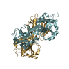
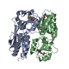
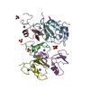
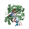
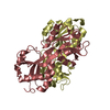
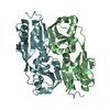
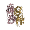
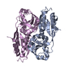
A: LYSR-TYPE REGULATORY PROTEIN B: LYSR-TYPE REGULATORY PROTEIN C: LYSR-TYPE REGULATORY PROTEIN D: LYSR-TYPE REGULATORY PROTEIN E: LYSR-TYPE REGULATORY PROTEIN F: LYSR-TYPE REGULATORY PROTEIN G: LYSR-TYPE REGULATORY PROTEIN H: LYSR-TYPE REGULATORY PROTEIN
Resolution: 2.99→43.9 Å / Cor.coef. Fo:Fc: 0.916 / Cor.coef. Fo:Fc free: 0.843 / SU B: 19.915 / SU ML: 0.373 / Cross valid method: THROUGHOUT / ESU R Free: 0.486 / Stereochemistry target values: MAXIMUM LIKELIHOOD / Details: HYDROGENS HAVE BEEN ADDED IN THE RIDING POSITIONS.
Rfactor
Num. reflection
% reflection
Selection details
Rfree
0.264
1883
5 %
RANDOM
Rwork
0.19364
-
-
-
obs
0.1972
35722
98.44 %
-
Solvent computation
Ion probe radii: 0.8 Å / Shrinkage radii: 0.8 Å / VDW probe radii: 1.4 Å / Solvent model: MASK
 Movie
Movie Controller


 Open data
 Basic information
 Components
 Keywords
 TRANSCRIPTION REGULATOR /
  Function and homology information
 BURKHOLDERIA SP. (bacteria)
 Authors
 Citation
 Structure visualization
 Downloads & links
 Other downloads
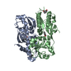


 PDBj
PDBj Assembly
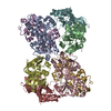
 Sample preparation
 / Beamline: ID23-2 / Wavelength: 0.873
  Processing