 Movie
Movie Controller

[English] 日本語
 Yorodumi
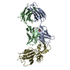
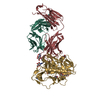
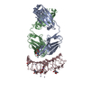
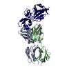
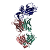
- PDB-2xwt: CRYSTAL STRUCTURE OF THE TSH RECEPTOR IN COMPLEX WITH A BLOCKING ... -
+ Open data

- Basic information
| Entry | Database: PDB / ID: 2xwt | |||||||||
|---|---|---|---|---|---|---|---|---|---|---|
| Title | CRYSTAL STRUCTURE OF THE TSH RECEPTOR IN COMPLEX WITH A BLOCKING TYPE TSHR AUTOANTIBODY | |||||||||
 Components Components |
| |||||||||
 Keywords Keywords |  SIGNALING PROTEIN/IMMUNE SYSTEM / SIGNALING PROTEIN-IMMUNE SYSTEM COMPLEX / GPCR / GRAVES' DISEASE / AUTOIMMUNITY / RECEPTOR-AUTOANTIBODY COMPLEX SIGNALING PROTEIN/IMMUNE SYSTEM / SIGNALING PROTEIN-IMMUNE SYSTEM COMPLEX / GPCR / GRAVES' DISEASE / AUTOIMMUNITY / RECEPTOR-AUTOANTIBODY COMPLEX | |||||||||
| Function / homology |  Function and homology information Function and homology informationthyroid-stimulating hormone signaling pathway / cellular response to glycoprotein / cellular response to thyrotropin-releasing hormone / thyroid-stimulating hormone receptor activity / Hormone ligand-binding receptors / G protein-coupled peptide receptor activity / G protein-coupled receptor signaling pathway, coupled to cyclic nucleotide second messenger / hormone-mediated signaling pathway / adenylate cyclase-activating G protein-coupled receptor signaling pathway / cell-cell signaling ...thyroid-stimulating hormone signaling pathway / cellular response to glycoprotein / cellular response to thyrotropin-releasing hormone / thyroid-stimulating hormone receptor activity / Hormone ligand-binding receptors / G protein-coupled peptide receptor activity / G protein-coupled receptor signaling pathway, coupled to cyclic nucleotide second messenger / hormone-mediated signaling pathway / adenylate cyclase-activating G protein-coupled receptor signaling pathway / cell-cell signaling / signaling receptor activity / positive regulation of cold-induced thermogenesis / G alpha (s) signalling events / basolateral plasma membrane / cell surface receptor signaling pathway / receptor complex / G protein-coupled receptor signaling pathway / positive regulation of cell population proliferation / protein-containing complex binding / cell surface / plasma membraneSimilarity search - Function | |||||||||
| Biological species |  HOMO SAPIENS (human) HOMO SAPIENS (human) | |||||||||
| Method | X-RAY DIFFRACTION / MOLECULAR REPLACEMENT / Resolution: 1.9 Å | |||||||||
 Authors Authors | Sanders, J. / Sanders, P. / Young, S. / Kabelis, K. / Baker, S. / Sullivan, A. / Evans, M. / Clark, J. / Wilmot, J. / Hu, X. ...Sanders, J. / Sanders, P. / Young, S. / Kabelis, K. / Baker, S. / Sullivan, A. / Evans, M. / Clark, J. / Wilmot, J. / Hu, X. / Roberts, E. / Powell, M. / Nunez Miguel, R. / Furmaniak, J. / Rees Smith, B. | |||||||||
 Citation Citation | Journal: J.Mol.Endocrinol. / Year: 2011 Title: Crystal Structure of the Tsh Receptor (Tshr) Bound to a Blocking-Type Tshr Autoantibody. Authors: Sanders, P. / Young, S. / Sanders, J. / Kabelis, K. / Baker, S. / Sullivan, A. / Evans, M. / Clark, J. / Wilmot, J. / Hu, X. / Roberts, E. / Powell, M. / Nunez Miguel, R. / Furmaniak, J. / Rees Smith, B. #1: Journal: Thyroid / Year: 2007Title: Crystal Structure of the Tsh Receptor in Complex with a Thyroid-Stimulating Autoantibody Authors: Sanders, J. / Chirgadze, D.Y. / Sanders, P. / Baker, S. / Sullivan, A. / Bhardwaja, A. / Bolton, J. / Reeve, M. / Nakatake, N. / Evans, M. / Richards, T. / Powell, M. / Miguel, R.N. / ...Authors: Sanders, J. / Chirgadze, D.Y. / Sanders, P. / Baker, S. / Sullivan, A. / Bhardwaja, A. / Bolton, J. / Reeve, M. / Nakatake, N. / Evans, M. / Richards, T. / Powell, M. / Miguel, R.N. / Blundell, T.L. / Furmaniak, J. / Smith, B.R. #2: Journal: Clinical Endocrinol. / Year: 2010 Title: Monoclonal Autoantibodies to the Tsh Receptor, One with Stimulating Activity and One with Blocking Activity, Obtained from the Same Blood Sample. Authors: Evans, M. / Sanders, J. / Tagami, T. / Sanders, P. / Young, S. / Roberts, E. / Wilmot, J. / Hu, X. / Kabelis, K. / Clark, J. / Holl, S. / Richards, T. / Collyer, A. / Furmaniak, J. / Smith, B.R. #3: Journal: Horm.Metab.Res. / Year: 2009 Title: Tsh Receptor - Autoantibody Interactions. Authors: Rees Smith, B. / Sanders, J. / Evans, M. / Tagami, T. / Furmaniak, J. #4: Journal: Lancet / Year: 2003 Title: Human Monoclonal Thyroid Stimulating Autoantibody Authors: Sanders, J. / Evans, M. / Premawardhana, L.D.K.E. / Depraetere, H. / Jeffreys, J. / Richards, T. / Furmaniak, J. / Smith, B.R. #5: Journal: Thyroid / Year: 2004 Title: Characteristics of a Human Monoclonal Autoantibody to the Thyrotropin Receptor: Sequence Structure and Function Authors: Sanders, J. / Jeffreys, J. / Depraetere, H. / Evans, M. / Richards, T. / Kiddie, A. / Brereton, K. / Premawardhana, L.D.K.E. / Chirgadze, D.Y. / Miguel, R.N. / Blundell, T.L. / Furmaniak, J. / Smith, B.R. #6: Journal: J.Mol.Endocrinol. / Year: 2009 Title: Thyroid Stimulating Autoantibody M22 Mimics Tsh Binding to the Tsh Receptor Leucine Rich Domain: A Comparative Structural Study of Protein-Protein Interactions. Authors: Nunez Miguel, R. / Sanders, J. / Chirgadze, D.Y. / Furmaniak, J. / Rees Smith, B. #7: Journal: Thyroid / Year: 2006 Title: Effects of Tsh Receptor Mutations on Binding and Biological Activity of Monoclonal Antibodies and Tsh Authors: Sanders, J. / Bolton, J. / Sanders, P. / Jeffreys, J. / Nakatake, N. / Richards, T. / Evans, M. / Kiddie, A. / Summerhayes, S. / Roberts, E. / Miguel, R.N. / Furmaniak, J. / Smith, B.R. #8: Journal: Thyroid / Year: 2007 Title: Molecular Interactions between the Tsh Receptor and a Thyroid-Stimulating Monoclonal Autoantibody Authors: Sanders, J. / Miguel, R.N. / Bolton, J. / Bhardwaja, A. / Sanders, P. / Nakatake, N. / Evans, M. / Furmaniak, J. / Smith, B.R. #9: Journal: J.Mol.Endocrinol. / Year: 2008 Title: Fsh and Tsh Binding to Their Respective Receptors: Similarities, Differences and Implication for Glycoprotein Hormone Specificity Authors: Miguel, R.N. / Sanders, J. / Chirgadze, D.Y. / Blundell, T.L. / Furmaniak, J. / Rees Smith, B. | |||||||||
| History |
|
- Structure visualization
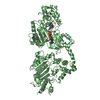
| Structure viewer | Molecule: MolmilJmol/JSmol |
|---|
- Downloads & links
-Download
| PDBx/mmCIF format | 2xwt.cif.gz | 164.6 KB | Display | PDBx/mmCIF format |
|---|---|---|---|---|
| PDB format | pdb2xwt.ent.gz | 125 KB | Display | PDB format |
| PDBx/mmJSON format | 2xwt.json.gz | Tree view | PDBx/mmJSON format | |
| Others |  Other downloads Other downloads |
-Validation report
| Arichive directory | https://data.pdbj.org/pub/pdb/validation_reports/xw/2xwtftp://data.pdbj.org/pub/pdb/validation_reports/xw/2xwt | HTTPS FTP |
|---|
-Related structure data
| Related structure data |  3g04S S: Starting model for refinement |
|---|---|
| Similar structure data |


-Links
 PDBj
PDBj


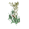
- Assembly
| Deposited unit | 
| ||||||||
|---|---|---|---|---|---|---|---|---|---|
| 1 |
| ||||||||
| Unit cell |
|
-Components
-Antibody , 2 types, 2 molecules AB
| #1: Antibody | Mass: 23637.561 Da / Num. of mol.: 1 / Fragment: FAB FRAGMENT HEAVY CHAIN, RESIDUES 1-229 Source method: isolated from a genetically manipulated source Source: (gene. exp.) HOMO SAPIENS (human)Production host: MOUSE-HUMAN HETEROHYBRIDOMA CELL LINE (others) |
|---|---|
| #2: Antibody | Mass: 22598.064 Da / Num. of mol.: 1 / Fragment: FAB FRAGMENT LIGHT CHAIN, RESIDUES 1-213 Source method: isolated from a genetically manipulated source Source: (gene. exp.) HOMO SAPIENS (human)Production host: MOUSE-HUMAN HETEROHYBRIDOMA CELL LINE (others) |
-Protein / Non-polymers , 2 types, 799 molecules C
| #3: Protein | / THYROID-STIMULATING HORMONE RECEPTOR / TSH-R Mass: 26943.828 Da / Num. of mol.: 1 / Fragment: LEUCINE RICH REPEAT DOMAIN, RESIDUES 22-260 Source method: isolated from a genetically manipulated source Source: (gene. exp.) HOMO SAPIENS (human) / Cell line (production host): High Five / Production host:  TRICHOPLUSIA NI (cabbage looper) / References: UniProt: P16473 TRICHOPLUSIA NI (cabbage looper) / References: UniProt: P16473 |
|---|---|
| #7: Water | ChemComp-HOH / WaterMass: 18.015 Da / Num. of mol.: 798 / Source method: isolated from a natural source / Formula: H2O |
-Sugars , 3 types, 4 molecules 
| #4: Polysaccharide | 2-acetamido-2-deoxy-beta-D-glucopyranose-(1-4)-2-acetamido-2-deoxy-beta-D-glucopyranose / Mass: 424.401 Da / Num. of mol.: 1 Source method: isolated from a genetically manipulated source |
|---|---|
| #5: Polysaccharide | alpha-D-mannopyranose-(1-6)-beta-D-mannopyranose-(1-4)-2-acetamido-2-deoxy-beta-D-glucopyranose-(1- ...alpha-D-mannopyranose-(1-6)-beta-D-mannopyranose-(1-4)-2-acetamido-2-deoxy-beta-D-glucopyranose-(1-4)-2-acetamido-2-deoxy-beta-D-glucopyranose / Mass: 748.682 Da / Num. of mol.: 1 Source method: isolated from a genetically manipulated source |
| #6: Sugar | N-Acetylglucosamine Type: D-saccharide, beta linking / Mass: 221.208 Da / Num. of mol.: 2 Type: D-saccharide, beta linking / Mass: 221.208 Da / Num. of mol.: 2Source method: isolated from a genetically manipulated source Formula: C8H15NO6 |
-Experimental details
-Experiment
| Experiment | Method: X-RAY DIFFRACTION / Number of used crystals: 1 |
|---|
- Sample preparation
| Crystal | Density Matthews: 2.62 Å3/Da / Density % sol: 53 % / Description: NONE |
|---|---|
| Crystal grow | pH: 5 / Details: 16% PEG 3350, 0.2 M SODIUM MALONATE PH 5. |
-Data collection
| Diffraction | Mean temperature: 100 K |
|---|---|
| Diffraction source | Source: ROTATING ANODE / Type: RIGAKU MICROMAX-007 HF / Wavelength: 1.54 |
| Detector | Type: RIGAKU RAXIS IV / Detector: IMAGE PLATE / Date: Nov 17, 2009 / Details: COLLIMATOR |
| Radiation | Protocol: SINGLE WAVELENGTH / Monochromatic (M) / Laue (L): M / Scattering type: x-ray |
| Radiation wavelength | Wavelength: 1.54 Å / Relative weight: 1 |
| Reflection | Resolution: 1.9→30 Å / Num. obs: 56573 / % possible obs: 94.5 % / Observed criterion σ(I): 0 / Redundancy: 5.1 % / Biso Wilson estimate: 0 Å2 / Rmerge(I) obs: 0.09 / Net I/σ(I): 11.7 |
| Reflection shell | Resolution: 1.9→1.95 Å / Rmerge(I) obs: 0.3 / Mean I/σ(I) obs: 3.5 / % possible all: 64.8 |
- Processing
| Software |
| ||||||||||||||||||||||||||||||||||||||||||||||||||||||||||||||||||||||||||||||||||||||||||||||||||||||||||||||||||||||||||||||||||||||||||||||||||||||||||||||||||||||||||||||||||||||
|---|---|---|---|---|---|---|---|---|---|---|---|---|---|---|---|---|---|---|---|---|---|---|---|---|---|---|---|---|---|---|---|---|---|---|---|---|---|---|---|---|---|---|---|---|---|---|---|---|---|---|---|---|---|---|---|---|---|---|---|---|---|---|---|---|---|---|---|---|---|---|---|---|---|---|---|---|---|---|---|---|---|---|---|---|---|---|---|---|---|---|---|---|---|---|---|---|---|---|---|---|---|---|---|---|---|---|---|---|---|---|---|---|---|---|---|---|---|---|---|---|---|---|---|---|---|---|---|---|---|---|---|---|---|---|---|---|---|---|---|---|---|---|---|---|---|---|---|---|---|---|---|---|---|---|---|---|---|---|---|---|---|---|---|---|---|---|---|---|---|---|---|---|---|---|---|---|---|---|---|---|---|---|---|
| Refinement | Method to determine structure: MOLECULAR REPLACEMENT Starting model: PDB ENTRY 3G04 Resolution: 1.9→66.98 Å / Cor.coef. Fo:Fc: 0.952 / Cor.coef. Fo:Fc free: 0.924 / SU B: 2.911 / SU ML: 0.087 / Cross valid method: THROUGHOUT / ESU R: 0.156 / ESU R Free: 0.149 / Stereochemistry target values: MAXIMUM LIKELIHOOD Details: HYDROGENS HAVE BEEN ADDED IN THE RIDING POSITIONS. U VALUES REFINED INDIVIDUALLY.
| ||||||||||||||||||||||||||||||||||||||||||||||||||||||||||||||||||||||||||||||||||||||||||||||||||||||||||||||||||||||||||||||||||||||||||||||||||||||||||||||||||||||||||||||||||||||
| Solvent computation | Ion probe radii: 0.8 Å / Shrinkage radii: 0.8 Å / VDW probe radii: 1.4 Å / Solvent model: MASK | ||||||||||||||||||||||||||||||||||||||||||||||||||||||||||||||||||||||||||||||||||||||||||||||||||||||||||||||||||||||||||||||||||||||||||||||||||||||||||||||||||||||||||||||||||||||
| Displacement parameters | Biso mean: 16.891 Å2
| ||||||||||||||||||||||||||||||||||||||||||||||||||||||||||||||||||||||||||||||||||||||||||||||||||||||||||||||||||||||||||||||||||||||||||||||||||||||||||||||||||||||||||||||||||||||
| Refinement step | Cycle: LAST / Resolution: 1.9→66.98 Å
| ||||||||||||||||||||||||||||||||||||||||||||||||||||||||||||||||||||||||||||||||||||||||||||||||||||||||||||||||||||||||||||||||||||||||||||||||||||||||||||||||||||||||||||||||||||||
| Refine LS restraints |
|