 Movie
Movie Controller

[English] 日本語
 Yorodumi
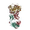
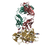
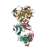
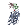
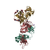
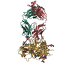
- PDB-6oz2: Crystal structure of the broadly neutralizing antibody N49P6 Fab ... -
+ Open data

- Basic information
| Entry | Database: PDB / ID: 6oz2 | ||||||||||||
|---|---|---|---|---|---|---|---|---|---|---|---|---|---|
| Title | Crystal structure of the broadly neutralizing antibody N49P6 Fab in complex with HIV-1 Clade A/E strain 93TH057 gp120 core. | ||||||||||||
 Components Components |
| ||||||||||||
 Keywords Keywords |  IMMUNE SYSTEM / HIV-1 / VRC01-CLASS ANTIBODY / CD4 BINDING SITE / CLADE A/E 93TH057 GP120 / VIRAL PROTEIN-IMMUNE SYSTEM COMPLEX / N49P6 IMMUNE SYSTEM / HIV-1 / VRC01-CLASS ANTIBODY / CD4 BINDING SITE / CLADE A/E 93TH057 GP120 / VIRAL PROTEIN-IMMUNE SYSTEM COMPLEX / N49P6 | ||||||||||||
| Function / homology | Gp120 core superfamily / Envelope glycoprotein GP120 / Human immunodeficiency virus 1, envelope glycoprotein Gp120 / viral envelope / clade A/E 93TH057 HIV-1 gp120 core Function and homology information Function and homology information | ||||||||||||
| Biological species |   Human immunodeficiency virus 1 Human immunodeficiency virus 1 Homo sapiens (human) Homo sapiens (human) | ||||||||||||
| Method | X-RAY DIFFRACTION / SYNCHROTRON / MOLECULAR REPLACEMENT / Resolution: 2.55 Å | ||||||||||||
 Authors Authors | Tolbert, W.D. / Pazgier, M. | ||||||||||||
| Funding support |  United States, 3items United States, 3items
| ||||||||||||
 Citation Citation | Journal: Mbio / Year: 2021 Title: Near-Pan-neutralizing, Plasma Deconvoluted Antibody N49P6 Mimics Host Receptor CD4 in Its Quaternary Interactions with the HIV-1 Envelope Trimer. Authors: Tolbert, W.D. / Nguyen, D.N. / Tehrani, Z.R. / Sajadi, M.M. / Pazgier, M. | ||||||||||||
| History |
|
- Structure visualization
| Structure viewer | Molecule: MolmilJmol/JSmol |
|---|
- Downloads & links
-Download
| PDBx/mmCIF format | 6oz2.cif.gz | 317.6 KB | Display | PDBx/mmCIF format |
|---|---|---|---|---|
| PDB format | pdb6oz2.ent.gz | 256.3 KB | Display | PDB format |
| PDBx/mmJSON format | 6oz2.json.gz | Tree view | PDBx/mmJSON format | |
| Others |  Other downloads Other downloads |
-Validation report
| Arichive directory | https://data.pdbj.org/pub/pdb/validation_reports/oz/6oz2ftp://data.pdbj.org/pub/pdb/validation_reports/oz/6oz2 | HTTPS FTP |
|---|
-Related structure data
| Related structure data |  6oz4C 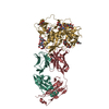 6bckS S: Starting model for refinement C: citing same article ( |
|---|---|
| Similar structure data |


-Links
 PDBj
PDBj


- Assembly
| Deposited unit | 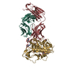
| ||||||||
|---|---|---|---|---|---|---|---|---|---|
| 1 |
| ||||||||
| Unit cell |
|
-Components
| #1: Protein | Mass: 39356.613 Da / Num. of mol.: 1 / Mutation: H375S Source method: isolated from a genetically manipulated source Source: (gene. exp.) Human immunodeficiency virus 1 / Gene: HIV-1 Env / Cell (production host): HEK 293 GnT1- / Production host: Homo sapiens (human) / References: UniProt: A0A0M3KKW9 | ||
|---|---|---|---|
| #2: Antibody | Mass: 24885.863 Da / Num. of mol.: 1 Source method: isolated from a genetically manipulated source Source: (gene. exp.) Homo sapiens (human) / Cell (production host): HEK 293 / Production host: Homo sapiens (human) | ||
| #3: Antibody | Mass: 21810.117 Da / Num. of mol.: 1 Source method: isolated from a genetically manipulated source Source: (gene. exp.) Homo sapiens (human) / Cell (production host): HEK 293 / Production host: Homo sapiens (human) | ||
| #4: Sugar | ChemComp-NAG / N-Acetylglucosamine  Type: D-saccharide, beta linking / Mass: 221.208 Da / Num. of mol.: 11 Type: D-saccharide, beta linking / Mass: 221.208 Da / Num. of mol.: 11Source method: isolated from a genetically manipulated source Formula: C8H15NO6 #5: Water | ChemComp-HOH / | Water Mass: 18.015 Da / Num. of mol.: 39 / Source method: isolated from a natural source / Formula: H2O Mass: 18.015 Da / Num. of mol.: 39 / Source method: isolated from a natural source / Formula: H2O |
-Experimental details
-Experiment
| Experiment | Method: X-RAY DIFFRACTION / Number of used crystals: 1 |
|---|
- Sample preparation
| Crystal | Density Matthews: 3 Å3/Da / Density % sol: 58.99 % |
|---|---|
| Crystal grow | Temperature: 294 K / Method: vapor diffusion, hanging drop / pH: 8 / Details: 15% PEG 3350 0.1 M Tris-HCl pH 8.0 |
-Data collection
| Diffraction | Mean temperature: 100 K / Serial crystal experiment: N |
|---|---|
| Diffraction source | Source: SYNCHROTRON / Site: SSRL / Beamline: BL12-2 / Wavelength: 0.97946 Å |
| Detector | Type: DECTRIS PILATUS 6M / Detector: PIXEL / Date: Nov 16, 2017 |
| Radiation | Monochromator: SI(111) / Protocol: SINGLE WAVELENGTH / Monochromatic (M) / Laue (L): M / Scattering type: x-ray |
| Radiation wavelength | Wavelength: 0.97946 Å / Relative weight: 1 |
| Reflection | Resolution: 2.55→50 Å / Num. obs: 31853 / % possible obs: 92 % / Redundancy: 3.1 % / Biso Wilson estimate: 40.8 Å2 / CC1/2: 0.99 / Rmerge(I) obs: 0.276 / Rpim(I) all: 0.166 / Net I/σ(I): 6.4 |
| Reflection shell | Resolution: 2.55→2.59 Å / Redundancy: 2.2 % / Rmerge(I) obs: 0.727 / Mean I/σ(I) obs: 1.1 / Num. unique obs: 1524 / CC1/2: 0.56 / Rpim(I) all: 0.556 / % possible all: 89.3 |
- Processing
| Software |
| ||||||||||||||||||||||||||||||||||||||||||||||||||||||||||||||||||||||||||||||||||||||||||||||||||||
|---|---|---|---|---|---|---|---|---|---|---|---|---|---|---|---|---|---|---|---|---|---|---|---|---|---|---|---|---|---|---|---|---|---|---|---|---|---|---|---|---|---|---|---|---|---|---|---|---|---|---|---|---|---|---|---|---|---|---|---|---|---|---|---|---|---|---|---|---|---|---|---|---|---|---|---|---|---|---|---|---|---|---|---|---|---|---|---|---|---|---|---|---|---|---|---|---|---|---|---|---|---|
| Refinement | Method to determine structure: MOLECULAR REPLACEMENT Starting model: 6BCK Resolution: 2.55→34.327 Å / SU ML: 0.43 / Cross valid method: FREE R-VALUE / σ(F): 1.33 / Phase error: 32.91
| ||||||||||||||||||||||||||||||||||||||||||||||||||||||||||||||||||||||||||||||||||||||||||||||||||||
| Solvent computation | Shrinkage radii: 0.9 Å / VDW probe radii: 1.11 Å | ||||||||||||||||||||||||||||||||||||||||||||||||||||||||||||||||||||||||||||||||||||||||||||||||||||
| Refinement step | Cycle: LAST / Resolution: 2.55→34.327 Å
| ||||||||||||||||||||||||||||||||||||||||||||||||||||||||||||||||||||||||||||||||||||||||||||||||||||
| Refine LS restraints |
| ||||||||||||||||||||||||||||||||||||||||||||||||||||||||||||||||||||||||||||||||||||||||||||||||||||
| LS refinement shell |
| ||||||||||||||||||||||||||||||||||||||||||||||||||||||||||||||||||||||||||||||||||||||||||||||||||||
| Refinement TLS params. | Method: refined / Refine-ID: X-RAY DIFFRACTION
| ||||||||||||||||||||||||||||||||||||||||||||||||||||||||||||||||||||||||||||||||||||||||||||||||||||
| Refinement TLS group |
|
