 Movie
Movie Controller

[English] 日本語
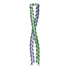
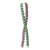
 Yorodumi
- PDB-2x7a: Structural basis of HIV-1 tethering to membranes by the Bst2-teth... -
+ Open data

- Basic information
| Entry | Database: PDB / ID: 2x7a | ||||||
|---|---|---|---|---|---|---|---|
| Title | Structural basis of HIV-1 tethering to membranes by the Bst2-tetherin ectodomain | ||||||
 Components Components | BONE MARROW STROMAL ANTIGEN 2 | ||||||
 Keywords Keywords |  IMMUNE SYSTEM / TETHERIN / GPI-ANCHOR / ANTIVIRAL DEFENSE / B-CELL ACTIVATION / SIGNAL-ANCHOR IMMUNE SYSTEM / TETHERIN / GPI-ANCHOR / ANTIVIRAL DEFENSE / B-CELL ACTIVATION / SIGNAL-ANCHOR | ||||||
| Function / homology |  Function and homology information Function and homology informationnegative regulation of plasmacytoid dendritic cell cytokine production / negative regulation of intracellular transport of viral material / response to interferon-beta / response to interferon-alpha / metalloendopeptidase inhibitor activity / positive regulation of leukocyte proliferation / azurophil granule membrane / negative regulation of viral genome replication / B cell activation / response to type II interferon ...negative regulation of plasmacytoid dendritic cell cytokine production / negative regulation of intracellular transport of viral material / response to interferon-beta / response to interferon-alpha / metalloendopeptidase inhibitor activity / positive regulation of leukocyte proliferation / azurophil granule membrane / negative regulation of viral genome replication / B cell activation / response to type II interferon / side of membrane / multivesicular body / negative regulation of cell migration / regulation of actin cytoskeleton organization / response to virus / negative regulation of cell growth / SARS-CoV-1 activates/modulates innate immune responses / Interferon alpha/beta signaling / positive regulation of canonical NF-kappaB signal transduction / defense response to virus / membrane raft / apical plasma membrane / intracellular membrane-bounded organelle / innate immune response / Neutrophil degranulation / Golgi apparatus / cell surface / protein homodimerization activity / RNA binding / extracellular exosome / membrane / identical protein binding / plasma membraneSimilarity search - Function | ||||||
| Biological species |  HOMO SAPIENS (human) HOMO SAPIENS (human) | ||||||
| Method | X-RAY DIFFRACTION / SYNCHROTRON / SAD / Resolution: 2.77 Å | ||||||
 Authors Authors | Natrajan, G. / McCarthy, A.A. / Weissenhorn, W. | ||||||
 Citation Citation | Journal: Cell Host Microbe / Year: 2010 Title: Structural Basis of HIV-1 Tethering to Membranes by the Bst-2/Tetherin Ectodomain. Authors: Hinz, A. / Miguet, N. / Natrajan, G. / Usami, Y. / Yamanaka, H. / Renesto, P. / Hartlieb, B. / Mccarthy, A.A. / Simorre, J.P. / Gottlinger, H. / Weissenhorn, W. | ||||||
| History |
|
- Structure visualization
| Structure viewer | Molecule: MolmilJmol/JSmol |
|---|
- Downloads & links
-Download
| PDBx/mmCIF format | 2x7a.cif.gz | 243.9 KB | Display | PDBx/mmCIF format |
|---|---|---|---|---|
| PDB format | pdb2x7a.ent.gz | 210.8 KB | Display | PDB format |
| PDBx/mmJSON format | 2x7a.json.gz | Tree view | PDBx/mmJSON format | |
| Others |  Other downloads Other downloads |
-Validation report
| Arichive directory | https://data.pdbj.org/pub/pdb/validation_reports/x7/2x7aftp://data.pdbj.org/pub/pdb/validation_reports/x7/2x7a | HTTPS FTP |
|---|
-Related structure data
| Similar structure data |
|---|


-Links
 PDBj
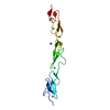
PDBj- Assembly
| Deposited unit | 
| ||||||||
|---|---|---|---|---|---|---|---|---|---|
| 1 | 
| ||||||||
| 2 | 
| ||||||||
| 3 | 
| ||||||||
| 4 | 
| ||||||||
| 5 | 
| ||||||||
| 6 | 
| ||||||||
| Unit cell |
|
-Components
-Protein , 1 types, 11 molecules ABCDEFGHIJK
| #1: Protein | Mass: 6894.455 Da / Num. of mol.: 11 / Fragment: RESIDUES 87-147 Source method: isolated from a genetically manipulated source Source: (gene. exp.) HOMO SAPIENS (human) / Production host:  ESCHERICHIA COLI (E. coli) / Strain (production host): ROSETTA2 / References: UniProt: Q10589 ESCHERICHIA COLI (E. coli) / Strain (production host): ROSETTA2 / References: UniProt: Q10589 |
|---|
-Non-polymers , 5 types, 76 molecules 


| #2: Chemical | ChemComp-NA /  Mass: 22.990 Da / Num. of mol.: 7 / Source method: obtained synthetically / Formula: Na Mass: 22.990 Da / Num. of mol.: 7 / Source method: obtained synthetically / Formula: Na#3: Chemical | ChemComp-CL / Chloride Mass: 35.453 Da / Num. of mol.: 7 / Source method: obtained synthetically / Formula: Cl Mass: 35.453 Da / Num. of mol.: 7 / Source method: obtained synthetically / Formula: Cl#4: Chemical | ChemComp-GOL / | Glycerol Mass: 92.094 Da / Num. of mol.: 1 / Source method: obtained synthetically / Formula: C3H8O3 Mass: 92.094 Da / Num. of mol.: 1 / Source method: obtained synthetically / Formula: C3H8O3#5: Chemical | ChemComp-MG / |  Mass: 24.305 Da / Num. of mol.: 1 / Source method: obtained synthetically / Formula: Mg Mass: 24.305 Da / Num. of mol.: 1 / Source method: obtained synthetically / Formula: Mg#6: Water | ChemComp-HOH / | WaterMass: 18.015 Da / Num. of mol.: 60 / Source method: isolated from a natural source / Formula: H2O |
|---|
-Experimental details
-Experiment
| Experiment | Method: X-RAY DIFFRACTION / Number of used crystals: 1 |
|---|
- Sample preparation
| Crystal | Density Matthews: 2.2 Å3/Da / Density % sol: 43.84 % / Description: NONE |
|---|---|
| Crystal grow | pH: 5 Details: 0.02M MGCL2, 0.1M BIS TRIS PH5.0, 20% POLYACRYLIC ACID |
-Data collection
| Diffraction | Mean temperature: 100 K |
|---|---|
| Diffraction source | Source: SYNCHROTRON / Site: ESRF  / Beamline: ID14-4 / Wavelength: 0.979 / Beamline: ID14-4 / Wavelength: 0.979 |
| Detector | Type: ADSC CCD / Detector: CCD / Date: Jun 1, 2009 / Details: MIRRORS |
| Radiation | Monochromator: CHANNEL CUT ESRF / Protocol: SINGLE WAVELENGTH / Monochromatic (M) / Laue (L): M / Scattering type: x-ray |
| Radiation wavelength | Wavelength: 0.979 Å / Relative weight: 1 |
| Reflection | Resolution: 2.77→45 Å / Num. obs: 35553 / % possible obs: 98.1 % / Observed criterion σ(I): 1.5 / Redundancy: 7.6 % / Biso Wilson estimate: 61.42 Å2 / Rmerge(I) obs: 0.09 / Net I/σ(I): 13.8 |
| Reflection shell | Resolution: 2.77→2.92 Å / Redundancy: 7.7 % / Rmerge(I) obs: 0.48 / Mean I/σ(I) obs: 3.8 / % possible all: 97.7 |
- Processing
| Software |
| ||||||||||||||||||||||||||||||||||||||||||||||||||||||||||||||||||||||||||||||||||||||||||||||||||||||||||||||||||||||||||||||||||||||||||||||||||||||||||||||||||||||||||||||||||||||||||||||||||||||||||||||||||||||||||||||||||||||||||||||||||||||||||||||||||||||||||||||||||||||||||||||||||||||||||||
|---|---|---|---|---|---|---|---|---|---|---|---|---|---|---|---|---|---|---|---|---|---|---|---|---|---|---|---|---|---|---|---|---|---|---|---|---|---|---|---|---|---|---|---|---|---|---|---|---|---|---|---|---|---|---|---|---|---|---|---|---|---|---|---|---|---|---|---|---|---|---|---|---|---|---|---|---|---|---|---|---|---|---|---|---|---|---|---|---|---|---|---|---|---|---|---|---|---|---|---|---|---|---|---|---|---|---|---|---|---|---|---|---|---|---|---|---|---|---|---|---|---|---|---|---|---|---|---|---|---|---|---|---|---|---|---|---|---|---|---|---|---|---|---|---|---|---|---|---|---|---|---|---|---|---|---|---|---|---|---|---|---|---|---|---|---|---|---|---|---|---|---|---|---|---|---|---|---|---|---|---|---|---|---|---|---|---|---|---|---|---|---|---|---|---|---|---|---|---|---|---|---|---|---|---|---|---|---|---|---|---|---|---|---|---|---|---|---|---|---|---|---|---|---|---|---|---|---|---|---|---|---|---|---|---|---|---|---|---|---|---|---|---|---|---|---|---|---|---|---|---|---|---|---|---|---|---|---|---|---|---|---|---|---|---|---|---|---|---|---|---|---|---|---|---|---|---|---|---|---|---|---|---|---|---|---|---|---|---|---|---|---|---|---|---|---|---|---|---|---|---|---|
| Refinement | Method to determine structure: SAD Starting model: NONE Resolution: 2.77→44.88 Å / SU ML: 0.44 / σ(F): 1.35 / Phase error: 28.46 / Stereochemistry target values: ML
| ||||||||||||||||||||||||||||||||||||||||||||||||||||||||||||||||||||||||||||||||||||||||||||||||||||||||||||||||||||||||||||||||||||||||||||||||||||||||||||||||||||||||||||||||||||||||||||||||||||||||||||||||||||||||||||||||||||||||||||||||||||||||||||||||||||||||||||||||||||||||||||||||||||||||||||
| Solvent computation | Shrinkage radii: 0.9 Å / VDW probe radii: 1.11 Å / Solvent model: FLAT BULK SOLVENT MODEL / Bsol: 62.917 Å2 / ksol: 0.323 e/Å3 | ||||||||||||||||||||||||||||||||||||||||||||||||||||||||||||||||||||||||||||||||||||||||||||||||||||||||||||||||||||||||||||||||||||||||||||||||||||||||||||||||||||||||||||||||||||||||||||||||||||||||||||||||||||||||||||||||||||||||||||||||||||||||||||||||||||||||||||||||||||||||||||||||||||||||||||
| Displacement parameters |
| ||||||||||||||||||||||||||||||||||||||||||||||||||||||||||||||||||||||||||||||||||||||||||||||||||||||||||||||||||||||||||||||||||||||||||||||||||||||||||||||||||||||||||||||||||||||||||||||||||||||||||||||||||||||||||||||||||||||||||||||||||||||||||||||||||||||||||||||||||||||||||||||||||||||||||||
| Refinement step | Cycle: LAST / Resolution: 2.77→44.88 Å
| ||||||||||||||||||||||||||||||||||||||||||||||||||||||||||||||||||||||||||||||||||||||||||||||||||||||||||||||||||||||||||||||||||||||||||||||||||||||||||||||||||||||||||||||||||||||||||||||||||||||||||||||||||||||||||||||||||||||||||||||||||||||||||||||||||||||||||||||||||||||||||||||||||||||||||||
| Refine LS restraints |
| ||||||||||||||||||||||||||||||||||||||||||||||||||||||||||||||||||||||||||||||||||||||||||||||||||||||||||||||||||||||||||||||||||||||||||||||||||||||||||||||||||||||||||||||||||||||||||||||||||||||||||||||||||||||||||||||||||||||||||||||||||||||||||||||||||||||||||||||||||||||||||||||||||||||||||||
| LS refinement shell |
| ||||||||||||||||||||||||||||||||||||||||||||||||||||||||||||||||||||||||||||||||||||||||||||||||||||||||||||||||||||||||||||||||||||||||||||||||||||||||||||||||||||||||||||||||||||||||||||||||||||||||||||||||||||||||||||||||||||||||||||||||||||||||||||||||||||||||||||||||||||||||||||||||||||||||||||
| Refinement TLS params. | Method: refined / Refine-ID: X-RAY DIFFRACTION
| ||||||||||||||||||||||||||||||||||||||||||||||||||||||||||||||||||||||||||||||||||||||||||||||||||||||||||||||||||||||||||||||||||||||||||||||||||||||||||||||||||||||||||||||||||||||||||||||||||||||||||||||||||||||||||||||||||||||||||||||||||||||||||||||||||||||||||||||||||||||||||||||||||||||||||||
| Refinement TLS group |
|
