 Movie
Movie Controller

[English] 日本語
 Yorodumi
- PDB-2wgg: Crystal Structure of Mycobacterium tuberculosis C171Q KasA varian... -
+ Open data

- Basic information
| Entry | Database: PDB / ID: 2wgg | ||||||
|---|---|---|---|---|---|---|---|
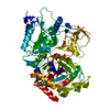
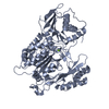
| Title | Crystal Structure of Mycobacterium tuberculosis C171Q KasA variant with bound TLM | ||||||
 Components Components | 3-OXOACYL-[ACYL-CARRIER-PROTEIN] SYNTHASE 1 | ||||||
 Keywords Keywords |  TRANSFERASE / BETA KETOACYL SYNTHASE I / CYTOPLASM / ACYLTRANSFERASE / LIPID SYNTHESIS / FATTY ACID BIOSYNTHESIS TRANSFERASE / BETA KETOACYL SYNTHASE I / CYTOPLASM / ACYLTRANSFERASE / LIPID SYNTHESIS / FATTY ACID BIOSYNTHESIS | ||||||
| Function / homology |  Function and homology information Function and homology informationmeromycolic acid 3-oxoacyl-(acyl carrier protein) synthase I / fatty acid elongation, saturated fatty acid / fatty acid elongation / acyltransferase activity / 3-oxoacyl-[acyl-carrier-protein] synthase activity / peptidoglycan-based cell wall / fatty acid biosynthetic process / identical protein binding / plasma membrane / cytosol / cytoplasmSimilarity search - Function | ||||||
| Biological species |   MYCOBACTERIUM TUBERCULOSIS (bacteria) MYCOBACTERIUM TUBERCULOSIS (bacteria) | ||||||
| Method | X-RAY DIFFRACTION / SYNCHROTRON / MOLECULAR REPLACEMENT / Resolution: 2 Å | ||||||
 Authors Authors | Luckner, S.R. / Kisker, C. | ||||||
 Citation Citation | Journal: Structure / Year: 2009 Title: Crystal Structures of Mycobacterium Tuberculosis Kasa Show Mode of Action within Cell Wall Biosynthesis and its Inhibition by Thiolactomycin Authors: Luckner, S.R. / Machutta, C.A. / Tonge, P.J. / Kisker, C. | ||||||
| History |
|
- Structure visualization
| Structure viewer | Molecule: MolmilJmol/JSmol |
|---|
- Downloads & links
-Download
| PDBx/mmCIF format | 2wgg.cif.gz | 621.7 KB | Display | PDBx/mmCIF format |
|---|---|---|---|---|
| PDB format | pdb2wgg.ent.gz | 516.4 KB | Display | PDB format |
| PDBx/mmJSON format | 2wgg.json.gz | Tree view | PDBx/mmJSON format | |
| Others |  Other downloads Other downloads |
-Validation report
| Arichive directory | https://data.pdbj.org/pub/pdb/validation_reports/wg/2wggftp://data.pdbj.org/pub/pdb/validation_reports/wg/2wgg | HTTPS FTP |
|---|
-Related structure data
| Related structure data | 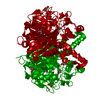 2wgdSC 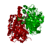 2wgeC 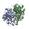 2wgfC C: citing same article ( S: Starting model for refinement |
|---|---|
| Similar structure data |


-Links
 PDBj
PDBj

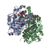
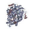
- Assembly
| Deposited unit | 
| ||||||||||||||||||||||||||||||||
|---|---|---|---|---|---|---|---|---|---|---|---|---|---|---|---|---|---|---|---|---|---|---|---|---|---|---|---|---|---|---|---|---|---|
| 1 | 
| ||||||||||||||||||||||||||||||||
| 2 | 
| ||||||||||||||||||||||||||||||||
| 3 | 
| ||||||||||||||||||||||||||||||||
| 4 | 
| ||||||||||||||||||||||||||||||||
| Unit cell |
| ||||||||||||||||||||||||||||||||
| Noncrystallographic symmetry (NCS) | NCS oper:
|
-Components
-Protein , 1 types, 8 molecules ABCDEFGH
| #1: Protein | Mass: 43384.969 Da / Num. of mol.: 8 / Mutation: YES Source method: isolated from a genetically manipulated source Source: (gene. exp.) MYCOBACTERIUM TUBERCULOSIS (bacteria) / Production host: MYCOBACTERIUM SMEGMATIS (bacteria) / Strain (production host): MC2155References: UniProt: P63454, UniProt: P9WQD9*PLUS, beta-ketoacyl-[acyl-carrier-protein] synthase I |
|---|
-Non-polymers , 5 types, 961 molecules 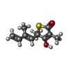


| #2: Chemical | ChemComp-TLM /  Mass: 210.293 Da / Num. of mol.: 8 / Source method: obtained synthetically / Formula: C11H14O2S Mass: 210.293 Da / Num. of mol.: 8 / Source method: obtained synthetically / Formula: C11H14O2S#3: Chemical | ChemComp-NA /  Mass: 22.990 Da / Num. of mol.: 8 / Source method: obtained synthetically / Formula: Na Mass: 22.990 Da / Num. of mol.: 8 / Source method: obtained synthetically / Formula: Na#4: Chemical | ChemComp-2PE / Polyethylene glycol Mass: 414.488 Da / Num. of mol.: 8 / Source method: obtained synthetically / Formula: C18H38O10 / Comment: precipitant*YM Mass: 414.488 Da / Num. of mol.: 8 / Source method: obtained synthetically / Formula: C18H38O10 / Comment: precipitant*YM#5: Chemical | ChemComp-PEG / Diethylene glycol Mass: 106.120 Da / Num. of mol.: 7 / Source method: obtained synthetically / Formula: C4H10O3 Mass: 106.120 Da / Num. of mol.: 7 / Source method: obtained synthetically / Formula: C4H10O3#6: Water | ChemComp-HOH / | WaterMass: 18.015 Da / Num. of mol.: 930 / Source method: isolated from a natural source / Formula: H2O |
|---|
-Details
| Compound details | ENGINEERED RESIDUE IN CHAIN A, CYS 171 TO GLN ENGINEERED RESIDUE IN CHAIN B, CYS 171 TO GLN ...ENGINEERED |
|---|---|
| Sequence details | CYS 171 IS MUTATED TO GLN 171 |
-Experimental details
-Experiment
| Experiment | Method: X-RAY DIFFRACTION / Number of used crystals: 1 |
|---|
- Sample preparation
| Crystal | Density Matthews: 2.84 Å3/Da / Density % sol: 56.74 % / Description: NONE |
|---|---|
| Crystal grow | Details: PEG3350, POTASSIUM FORMATE |
-Data collection
| Diffraction | Mean temperature: 100 K |
|---|---|
| Diffraction source | Source: SYNCHROTRON / Site: BESSY  / Beamline: 14.2 / Wavelength: 1 / Beamline: 14.2 / Wavelength: 1 |
| Detector | Type: MARMOSAIC 225 mm CCD / Detector: CCD |
| Radiation | Protocol: SINGLE WAVELENGTH / Monochromatic (M) / Laue (L): M / Scattering type: x-ray |
| Radiation wavelength | Wavelength: 1 Å / Relative weight: 1 |
| Reflection twin | Operator: h,-h-k,-l / Fraction: 0.287 |
| Reflection | Resolution: 2→46.18 Å / Num. obs: 996668 / % possible obs: 100 % / Observed criterion σ(I): 1 / Redundancy: 3.89 % / Biso Wilson estimate: 23.8 Å2 / Rmerge(I) obs: 0.07 / Net I/σ(I): 14.35 |
| Reflection shell | Resolution: 2→2.11 Å / Redundancy: 3.85 % / Rmerge(I) obs: 0.35 / Mean I/σ(I) obs: 3.8 / % possible all: 100 |
- Processing
| Software |
| |||||||||||||||||||||||||||||||||||||||||||||||||||||||||||||||||||||||||||||||||||||||||||||||||||||||||||||||||||||||||||||||||||||||||||||||||||
|---|---|---|---|---|---|---|---|---|---|---|---|---|---|---|---|---|---|---|---|---|---|---|---|---|---|---|---|---|---|---|---|---|---|---|---|---|---|---|---|---|---|---|---|---|---|---|---|---|---|---|---|---|---|---|---|---|---|---|---|---|---|---|---|---|---|---|---|---|---|---|---|---|---|---|---|---|---|---|---|---|---|---|---|---|---|---|---|---|---|---|---|---|---|---|---|---|---|---|---|---|---|---|---|---|---|---|---|---|---|---|---|---|---|---|---|---|---|---|---|---|---|---|---|---|---|---|---|---|---|---|---|---|---|---|---|---|---|---|---|---|---|---|---|---|---|---|---|---|
| Refinement | Method to determine structure: MOLECULAR REPLACEMENT Starting model: PDB ENTRY 2WGD Resolution: 2→46.16 Å / σ(F): 1.08 / Phase error: 26.89 / Stereochemistry target values: TWIN_LSQ_F
| |||||||||||||||||||||||||||||||||||||||||||||||||||||||||||||||||||||||||||||||||||||||||||||||||||||||||||||||||||||||||||||||||||||||||||||||||||
| Solvent computation | Shrinkage radii: 0.9 Å / VDW probe radii: 1.11 Å / Solvent model: FLAT BULK SOLVENT MODEL / Bsol: 33.68 Å2 / ksol: 0.37 e/Å3 | |||||||||||||||||||||||||||||||||||||||||||||||||||||||||||||||||||||||||||||||||||||||||||||||||||||||||||||||||||||||||||||||||||||||||||||||||||
| Displacement parameters | Biso mean: 29.3 Å2
| |||||||||||||||||||||||||||||||||||||||||||||||||||||||||||||||||||||||||||||||||||||||||||||||||||||||||||||||||||||||||||||||||||||||||||||||||||
| Refinement step | Cycle: LAST / Resolution: 2→46.16 Å
| |||||||||||||||||||||||||||||||||||||||||||||||||||||||||||||||||||||||||||||||||||||||||||||||||||||||||||||||||||||||||||||||||||||||||||||||||||
| Refine LS restraints |
| |||||||||||||||||||||||||||||||||||||||||||||||||||||||||||||||||||||||||||||||||||||||||||||||||||||||||||||||||||||||||||||||||||||||||||||||||||
| LS refinement shell |
|
