[English] 日本語
Yorodumi
- PDB-2w9a: Ternary complex of Dpo4 bound to N2,N2-dimethyl-deoxyguanosine mo... -

+
Open data


ID or keywords:

Loading...

-
Basic information

Entry
Database: PDB / ID: 2w9a
TitleTernary complex of Dpo4 bound to N2,N2-dimethyl-deoxyguanosine modified DNA with incoming dGTP
Components
  • 5'-D(*GP*GP*GP*GP*GP*AP*AP*GP*GP*AP *TP*TP*CP*DOC)-3'
  • 5'-D(*TP*CP*AP*CP*O2GP*GP*AP*AP*TP*CP *CP*TP*TP*CP*CP*CP*CP*C)-3'
  • DNA POLYMERASE IV
Keywords
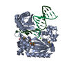
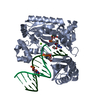
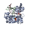
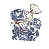
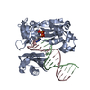
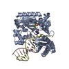
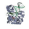
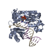
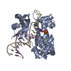
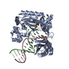
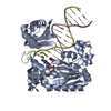
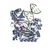
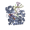
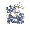
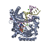
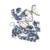
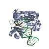
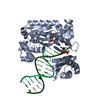
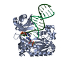
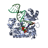
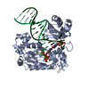
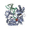
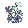
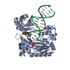
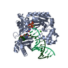
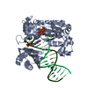
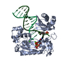
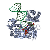
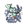
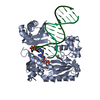
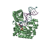
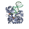
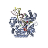
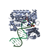
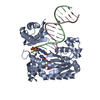
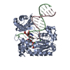
TRANSFERASE/DNA / TRANSFERASE DNA COMPLEX / N2-DIMETHYL-G / METAL-BINDING / MUTATOR PROTEIN / NUCLEOTIDYLTRANSFERASE / DNA-DIRECTED DNA POLYMERASE / DNA DAMAGE / DNA REPAIR / DNA-BINDING / TRANSFERASE / DNA REPLICATION / DNA / DPO4 / ADDUCT / CYTOPLASM / MAGNESIUM / POLYMERASE / TRANSFERASE-DNA complex
Function / homology
Function and homology information


SOS response / error-prone translesion synthesis / DNA-templated DNA replication / damaged DNA binding / DNA-directed DNA polymerase / DNA-directed DNA polymerase activity / magnesium ion binding / cytosol
Similarity search - Function
DNA polymerase type-Y, HhH motif / IMS family HHH motif / DNA polymerase IV / DNA polymerase, Y-family, little finger domain / MutS, DNA mismatch repair protein, domain I - #60 / MutS, DNA mismatch repair protein, domain I / DNA polymerase, Y-family, little finger domain / impB/mucB/samB family C-terminal domain / UmuC domain / DNA polymerase, Y-family, little finger domain superfamily ...DNA polymerase type-Y, HhH motif / IMS family HHH motif / DNA polymerase IV / DNA polymerase, Y-family, little finger domain / MutS, DNA mismatch repair protein, domain I - #60 / MutS, DNA mismatch repair protein, domain I / DNA polymerase, Y-family, little finger domain / impB/mucB/samB family C-terminal domain / UmuC domain / DNA polymerase, Y-family, little finger domain superfamily / impB/mucB/samB family / UmuC domain profile. / Reverse transcriptase/Diguanylate cyclase domain / Dna Ligase; domain 1 / 5' to 3' exonuclease, C-terminal subdomain / DNA polymerase; domain 1 / Reverse transcriptase/Diguanylate cyclase domain / Alpha-Beta Plaits / DNA/RNA polymerase superfamily / 2-Layer Sandwich / Orthogonal Bundle / 3-Layer(aba) Sandwich / Mainly Alpha / Alpha Beta
Similarity search - Domain/homology
2'-DEOXYGUANOSINE-5'-TRIPHOSPHATE / DNA / DNA (> 10) / DNA polymerase IV
Similarity search - Component
Biological speciesSULFOLOBUS SOLFATARICUS (archaea)
MethodX-RAY DIFFRACTION / SYNCHROTRON / MOLECULAR REPLACEMENT / Resolution: 2.6 Å
AuthorsEoff, R.L. / Zhang, H. / Kosekov, I.D. / Rizzo, C.J. / Egli, M. / Guengerich, F.P.
CitationJournal: J.Biol.Chem. / Year: 2009
Title: Structure-Function Relationships in Miscoding by Sulfolobus Solfataricus DNA Polymerase Dpo4: Guanine N2,N2-Dimethyl Substitution Produces Inactive and Miscoding Polymerase Complexes.
Authors: Zhang, H. / Eoff, R.L. / Kozekov, I.D. / Rizzo, C.J. / Egli, M. / Guengerich, F.P.
History
DepositionJan 22, 2009Deposition site: PDBE / Processing site: PDBE
Revision 1.0May 12, 2009Provider: repository / Type: Initial release
Revision 1.1May 8, 2011Group: Version format compliance
Revision 1.2Jul 13, 2011Group: Version format compliance
Revision 1.3Dec 13, 2023Group: Data collection / Database references ...Data collection / Database references / Derived calculations / Other / Refinement description
Category: chem_comp_atom / chem_comp_bond ...chem_comp_atom / chem_comp_bond / database_2 / pdbx_database_status / pdbx_initial_refinement_model / pdbx_struct_conn_angle / struct_conn / struct_conn_type / struct_site
Item: _database_2.pdbx_DOI / _database_2.pdbx_database_accession ..._database_2.pdbx_DOI / _database_2.pdbx_database_accession / _pdbx_database_status.status_code_sf / _pdbx_struct_conn_angle.ptnr1_auth_asym_id / _pdbx_struct_conn_angle.ptnr1_auth_comp_id / _pdbx_struct_conn_angle.ptnr1_auth_seq_id / _pdbx_struct_conn_angle.ptnr1_label_asym_id / _pdbx_struct_conn_angle.ptnr1_label_atom_id / _pdbx_struct_conn_angle.ptnr1_label_comp_id / _pdbx_struct_conn_angle.ptnr1_label_seq_id / _pdbx_struct_conn_angle.ptnr3_auth_asym_id / _pdbx_struct_conn_angle.ptnr3_auth_comp_id / _pdbx_struct_conn_angle.ptnr3_auth_seq_id / _pdbx_struct_conn_angle.ptnr3_label_asym_id / _pdbx_struct_conn_angle.ptnr3_label_atom_id / _pdbx_struct_conn_angle.ptnr3_label_comp_id / _pdbx_struct_conn_angle.ptnr3_label_seq_id / _pdbx_struct_conn_angle.value / _struct_conn.conn_type_id / _struct_conn.id / _struct_conn.pdbx_dist_value / _struct_conn.pdbx_leaving_atom_flag / _struct_conn.ptnr1_auth_asym_id / _struct_conn.ptnr1_auth_comp_id / _struct_conn.ptnr1_auth_seq_id / _struct_conn.ptnr1_label_asym_id / _struct_conn.ptnr1_label_atom_id / _struct_conn.ptnr1_label_comp_id / _struct_conn.ptnr1_label_seq_id / _struct_conn.ptnr2_auth_asym_id / _struct_conn.ptnr2_auth_comp_id / _struct_conn.ptnr2_auth_seq_id / _struct_conn.ptnr2_label_asym_id / _struct_conn.ptnr2_label_atom_id / _struct_conn.ptnr2_label_comp_id / _struct_conn.ptnr2_label_seq_id / _struct_conn_type.id / _struct_site.pdbx_auth_asym_id / _struct_site.pdbx_auth_comp_id / _struct_site.pdbx_auth_seq_id

-
Structure visualization

Structure viewerMolecule:
MolmilJmol/JSmol

Downloads & links

-
Assembly

Deposited unit
A: DNA POLYMERASE IV
P: 5'-D(*GP*GP*GP*GP*GP*AP*AP*GP*GP*AP *TP*TP*CP*DOC)-3'
T: 5'-D(*TP*CP*AP*CP*O2GP*GP*AP*AP*TP*CP *CP*TP*TP*CP*CP*CP*CP*C)-3'
hetero molecules


Theoretical massNumber of molelcules
Total (without water)51,4628
Polymers50,8573
Non-polymers6045
Water1,17165
1


  • Idetical with deposited unit
  • defined by author&software
TypeNameSymmetry operationNumber
identity operation1_555x,y,z1
Buried area5780 Å2
ΔGint-69 kcal/mol
Surface area20260 Å2
MethodPISA
Unit cell
Length a, b, c (Å)94.670, 103.757, 52.750
Angle α, β, γ (deg.)90.00, 90.00, 90.00
Int Tables number18
Space group name H-MP21212

-
Components

-
Protein , 1 types, 1 molecules A

#1: Protein DNA POLYMERASE IV / / POL IV / DPO4


Mass: 41086.754 Da / Num. of mol.: 1
Source method: isolated from a genetically manipulated source
Details: Y-FAMILY DNA POLYMERASE FROM SULFOLOBUS SOLFATARICUS
Source: (gene. exp.) SULFOLOBUS SOLFATARICUS (archaea) / Strain: P2 / Plasmid: PET22B / Production host: ESCHERICHIA COLI (E. coli) / Strain (production host): BL21(DE3) / References: UniProt: Q97W02, DNA-directed DNA polymerase

-
DNA chain , 2 types, 2 molecules PT

#2: DNA chain 5'-D(*GP*GP*GP*GP*GP*AP*AP*GP*GP*AP *TP*TP*CP*DOC)-3' / PRIMER DNA


Mass: 4369.854 Da / Num. of mol.: 1 / Source method: obtained synthetically / Details: 14 BASE PRIMER DNA 5'-GGG GGA AGG ATT C(DOC) -3'
#3: DNA chain 5'-D(*TP*CP*AP*CP*O2GP*GP*AP*AP*TP*CP *CP*TP*TP*CP*CP*CP*CP*C)-3' / TEMPLATE DNA


Mass: 5400.534 Da / Num. of mol.: 1 / Source method: obtained synthetically
Details: 18 BASE TEMPLATE DNA 5'-TCA C(O2G)G AAT CCT TCC CCC-3'

-
Non-polymers , 3 types, 70 molecules

#4: Chemical ChemComp-DGT / 2'-DEOXYGUANOSINE-5'-TRIPHOSPHATE / Deoxyguanosine triphosphate


Mass: 507.181 Da / Num. of mol.: 1 / Source method: obtained synthetically / Formula: C10H16N5O13P3
#5: Chemical
ChemComp-MG / MAGNESIUM ION


Mass: 24.305 Da / Num. of mol.: 4 / Source method: obtained synthetically / Formula: Mg
#6: Water ChemComp-HOH / water / Water


Mass: 18.015 Da / Num. of mol.: 65 / Source method: isolated from a natural source / Formula: H2O

-
Details

Sequence detailsENGINEERED HEXA-HISTIDINE TAG AND LAST 11 RESIDUES ARE DISORDERED

-
Experimental details

-
Experiment

ExperimentMethod: X-RAY DIFFRACTION / Number of used crystals: 1

-
Sample preparation

CrystalDensity Matthews: 2.44 Å3/Da / Density % sol: 53.74 % / Description: NONE
Crystal growpH: 7.4
Details: DPO4\: DNA COMPLEX (1\:1.2 MOLAR RATIO, 150 MICROMOLAR), 20 MM TRIS HCL (PH 7.4), 15% POLYETHYLENE GLYCOL 3350 (W/V), 60 MM SODIUM CHLORIDE, 5 MM MAGNESIUM CHLORIDE, 1 MM DGTP.

-
Data collection

DiffractionMean temperature: 110 K
Diffraction sourceSource: SYNCHROTRON / Site: APS / Beamline: 21-ID / Wavelength: 0.97857
DetectorType: MARRESEARCH / Detector: CCD / Date: Jun 22, 2008 / Details: MIRRORS
RadiationMonochromator: SI(111) / Protocol: SINGLE WAVELENGTH / Monochromatic (M) / Laue (L): M / Scattering type: x-ray
Radiation wavelengthWavelength: 0.97857 Å / Relative weight: 1
ReflectionResolution: 2.55→19.94 Å / Num. obs: 16285 / % possible obs: 98.2 % / Observed criterion σ(I): 0 / Redundancy: 6.8 % / Biso Wilson estimate: 42.4 Å2 / Rmerge(I) obs: 0.06 / Net I/σ(I): 23.3
Reflection shellResolution: 2.55→2.69 Å / Redundancy: 6.2 % / Rmerge(I) obs: 0.2 / Mean I/σ(I) obs: 5.3 / % possible all: 97.4

-
Processing

Software
NameVersionClassification
CNS1.2refinement
XDSdata reduction
XDSdata scaling
MOLREPphasing
RefinementMethod to determine structure: MOLECULAR REPLACEMENT
Starting model: PDB ENTRY 2JEF
Resolution: 2.6→19.94 Å / Rfactor Rfree error: 0.009 / Data cutoff high absF: 68406.28 / Data cutoff low absF: 0 / Isotropic thermal model: RESTRAINED / Cross valid method: THROUGHOUT / σ(F): 0 / Details: BULK SOLVENT MODEL USED
RfactorNum. reflection% reflectionSelection details
Rfree0.246 790 4.9 %RANDOM
Rwork0.213 ---
obs0.213 16285 98.3 %-
Solvent computationSolvent model: FLAT MODEL / Bsol: 31.36 Å2 / ksol: 0.35 e/Å3
Displacement parametersBiso mean: 42.7 Å2
Baniso -1Baniso -2Baniso -3
1-5.69 Å20 Å20 Å2
2---3.13 Å20 Å2
3----2.56 Å2
Refine analyze
FreeObs
Luzzati coordinate error0.4 Å0.33 Å
Luzzati d res low-30 Å
Luzzati sigma a0.4 Å0.35 Å
Refinement stepCycle: LAST / Resolution: 2.6→19.94 Å
ProteinNucleic acidLigandSolventTotal
Num. atoms2754 609 35 65 3463
Refine LS restraints
Refine-IDTypeDev idealDev ideal target
X-RAY DIFFRACTIONc_bond_d0.009
X-RAY DIFFRACTIONc_bond_d_na
X-RAY DIFFRACTIONc_bond_d_prot
X-RAY DIFFRACTIONc_angle_d
X-RAY DIFFRACTIONc_angle_d_na
X-RAY DIFFRACTIONc_angle_d_prot
X-RAY DIFFRACTIONc_angle_deg1.4
X-RAY DIFFRACTIONc_angle_deg_na
X-RAY DIFFRACTIONc_angle_deg_prot
X-RAY DIFFRACTIONc_dihedral_angle_d21.9
X-RAY DIFFRACTIONc_dihedral_angle_d_na
X-RAY DIFFRACTIONc_dihedral_angle_d_prot
X-RAY DIFFRACTIONc_improper_angle_d1.83
X-RAY DIFFRACTIONc_improper_angle_d_na
X-RAY DIFFRACTIONc_improper_angle_d_prot
X-RAY DIFFRACTIONc_mcbond_it1.231.5
X-RAY DIFFRACTIONc_mcangle_it2.122
X-RAY DIFFRACTIONc_scbond_it1.852
X-RAY DIFFRACTIONc_scangle_it2.952.5
Refine LS restraints NCSNCS model details: NONE
LS refinement shellResolution: 2.55→2.71 Å / Rfactor Rfree error: 0.036 / Total num. of bins used: 6
RfactorNum. reflection% reflection
Rfree0.336 85 4.6 %
Rwork0.28 1767 -
obs--64.6 %
Xplor file
Refine-IDSerial noParam fileTopol file
X-RAY DIFFRACTION1PROTEIN_REP.PARAMPROTEIN.TOP
X-RAY DIFFRACTION2DNA-RNA_REP.PARAMDNA-RNA.TOP
X-RAY DIFFRACTION3DGTP.PARAMDGTP.TOP
X-RAY DIFFRACTION4ION.PARAMION.TOP
X-RAY DIFFRACTION5WATER_REP.PARAMWATER.TOP

+
About Yorodumi

-
News

-
Feb 9, 2022. New format data for meta-information of EMDB entries

New format data for meta-information of EMDB entries

  • Version 3 of the EMDB header file is now the official format.
  • The previous official version 1.9 will be removed from the archive.

Related info.:EMDB header

External links:wwPDB to switch to version 3 of the EMDB data model

-
Aug 12, 2020. Covid-19 info

Covid-19 info

URL: https://pdbj.org/emnavi/covid19.php

New page: Covid-19 featured information page in EM Navigator.

Related info.:Covid-19 info / Mar 5, 2020. Novel coronavirus structure data

+
Mar 5, 2020. Novel coronavirus structure data

Novel coronavirus structure data

Related info.:Yorodumi Speices / Aug 12, 2020. Covid-19 info

External links:COVID-19 featured content - PDBj / Molecule of the Month (242):Coronavirus Proteases

+
Jan 31, 2019. EMDB accession codes are about to change! (news from PDBe EMDB page)

EMDB accession codes are about to change! (news from PDBe EMDB page)

  • The allocation of 4 digits for EMDB accession codes will soon come to an end. Whilst these codes will remain in use, new EMDB accession codes will include an additional digit and will expand incrementally as the available range of codes is exhausted. The current 4-digit format prefixed with “EMD-” (i.e. EMD-XXXX) will advance to a 5-digit format (i.e. EMD-XXXXX), and so on. It is currently estimated that the 4-digit codes will be depleted around Spring 2019, at which point the 5-digit format will come into force.
  • The EM Navigator/Yorodumi systems omit the EMD- prefix.

Related info.:Q: What is EMD? / ID/Accession-code notation in Yorodumi/EM Navigator

External links:EMDB Accession Codes are Changing Soon! / Contact to PDBj

+
Jul 12, 2017. Major update of PDB

Major update of PDB

  • wwPDB released updated PDB data conforming to the new PDBx/mmCIF dictionary.
  • This is a major update changing the version number from 4 to 5, and with Remediation, in which all the entries are updated.
  • In this update, many items about electron microscopy experimental information are reorganized (e.g. em_software).
  • Now, EM Navigator and Yorodumi are based on the updated data.

External links:wwPDB Remediation / Enriched Model Files Conforming to OneDep Data Standards Now Available in the PDB FTP Archive

-
Yorodumi

Thousand views of thousand structures

  • Yorodumi is a browser for structure data from EMDB, PDB, SASBDB, etc.
  • This page is also the successor to EM Navigator detail page, and also detail information page/front-end page for Omokage search.
  • The word "yorodu" (or yorozu) is an old Japanese word meaning "ten thousand". "mi" (miru) is to see.

Related info.:EMDB / PDB / SASBDB / Comparison of 3 databanks / Yorodumi Search / Aug 31, 2016. New EM Navigator & Yorodumi / Yorodumi Papers / Jmol/JSmol / Function and homology information / Changes in new EM Navigator and Yorodumi

Read more