 Movie
Movie Controller

[English] 日本語
 Yorodumi
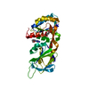
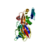
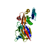
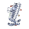
- PDB-2rsp: STRUCTURE OF THE ASPARTIC PROTEASE FROM ROUS SARCOMA RETROVIRUS R... -
+ Open data

- Basic information
| Entry | Database: PDB / ID: 2rsp | ||||||
|---|---|---|---|---|---|---|---|
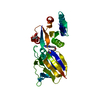
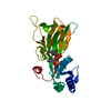
| Title | STRUCTURE OF THE ASPARTIC PROTEASE FROM ROUS SARCOMA RETROVIRUS REFINED AT 2 ANGSTROMS RESOLUTION | ||||||
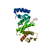
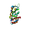
 Components Components | RSV PROTEASE | ||||||
 Keywords Keywords | HYDROLASE(ASPARTYL PROTEINASE) | ||||||
| Function / homology |  Function and homology information Function and homology informationhost cell nucleoplasm / viral procapsid maturation / host cell nucleolus /  Hydrolases; Acting on peptide bonds (peptidases); Aspartic endopeptidases / viral capsid / structural constituent of virion / nucleic acid binding / aspartic-type endopeptidase activity / host cell plasma membrane / proteolysis ...host cell nucleoplasm / viral procapsid maturation / host cell nucleolus / Hydrolases; Acting on peptide bonds (peptidases); Aspartic endopeptidases / viral capsid / structural constituent of virion / nucleic acid binding / aspartic-type endopeptidase activity / host cell plasma membrane / proteolysis / zinc ion binding / membrane Hydrolases; Acting on peptide bonds (peptidases); Aspartic endopeptidases / viral capsid / structural constituent of virion / nucleic acid binding / aspartic-type endopeptidase activity / host cell plasma membrane / proteolysis ...host cell nucleoplasm / viral procapsid maturation / host cell nucleolus / Hydrolases; Acting on peptide bonds (peptidases); Aspartic endopeptidases / viral capsid / structural constituent of virion / nucleic acid binding / aspartic-type endopeptidase activity / host cell plasma membrane / proteolysis / zinc ion binding / membraneSimilarity search - Function | ||||||
| Biological species |  Rous sarcoma virus Rous sarcoma virus | ||||||
| Method | X-RAY DIFFRACTION / Resolution: 2 Å | ||||||
 Authors Authors | Wlodawer, A. / Miller, M. / Jaskolski, M. | ||||||
 Citation Citation | Journal: Biochemistry / Year: 1990 Title: Structure of the aspartic protease from Rous sarcoma retrovirus refined at 2-A resolution. Authors: Jaskolski, M. / Miller, M. / Rao, J.K. / Leis, J. / Wlodawer, A. #1: Journal: Nature / Year: 1989Title: Crystal Structure of a Retroviral Protease Proves Relationship to Aspartic Protease Family Authors: Miller, M. / Jaskolski, M. / Rao, J.K.M. / Leis, J. / Wlodawer, A. | ||||||
| History |
| ||||||
| Remark 700 | SHEET THE DIMER INTERFACE IS COMPOSED OF INTERDIGITATED N- AND C-TERMINI FROM BOTH SUBUNITS FORMING ...SHEET THE DIMER INTERFACE IS COMPOSED OF INTERDIGITATED N- AND C-TERMINI FROM BOTH SUBUNITS FORMING A FOUR-STRANDED ANTIPARALLEL BETA-SHEET (SHEET *INT* BELOW). SHEETS MA AND MB ARE EACH DISRUPTED BY BULGES AT RESIDUES ASP 78 IN STRAND 2 AND ARG 93 IN STRAND 3. |
- Structure visualization
| Structure viewer | Molecule: MolmilJmol/JSmol |
|---|
- Downloads & links
-Download
| PDBx/mmCIF format | 2rsp.cif.gz | 62.5 KB | Display | PDBx/mmCIF format |
|---|---|---|---|---|
| PDB format | pdb2rsp.ent.gz | 45.9 KB | Display | PDB format |
| PDBx/mmJSON format | 2rsp.json.gz | Tree view | PDBx/mmJSON format | |
| Others |  Other downloads Other downloads |
-Validation report
| Arichive directory | https://data.pdbj.org/pub/pdb/validation_reports/rs/2rspftp://data.pdbj.org/pub/pdb/validation_reports/rs/2rsp | HTTPS FTP |
|---|
-Related structure data
| Similar structure data |
|---|


-Links
 PDBj
PDBj- Assembly
| Deposited unit | 
| |||||||||
|---|---|---|---|---|---|---|---|---|---|---|
| 1 |
| |||||||||
| Unit cell |
| |||||||||
| Components on special symmetry positions |
| |||||||||
| Details | THE ACTIVE ENZYME IS A DIMER COMPOSED OF TWO RSV PR MOLECULES. THIS DIMER COMPRISES THE CRYSTALLOGRAPHIC ASYMMETRIC UNIT. THE MOLECULES IN THIS DIMER ARE RELATED BY A NON-CRYSTALLOGRAPHIC TWO-FOLD AXIS WHICH IS NEARLY COINCIDENT WITH THE (2 1 -3) CRYSTAL DIRECTION. CHAIN IDENTIFIERS *A* AND *B* HAVE BEEN ASSIGNED TO THE TWO MOLECULES PRESENTED IN THIS ENTRY. |
-Components
| #1: Protein | Mass: 13625.862 Da / Num. of mol.: 2 Source method: isolated from a genetically manipulated source Source: (gene. exp.) Rous sarcoma virus / Genus: Alpharetrovirus / References: UniProt: P03322#2: Water | ChemComp-HOH / | Water Mass: 18.015 Da / Num. of mol.: 252 / Source method: isolated from a natural source / Formula: H2O Mass: 18.015 Da / Num. of mol.: 252 / Source method: isolated from a natural source / Formula: H2OCompound details | THE CATALYTIC SITE IS FORMED BY TWO ASP-SER-GLY LOOPS, ONE FORM EACH SUBUNIT, CONNECTED BY AN ...THE CATALYTIC SITE IS FORMED BY TWO ASP-SER-GLY LOOPS, ONE FORM EACH SUBUNIT, CONNECTED BY AN INTRICATE SYSTEM OF HYDROGEN BONDS. | |
|---|
-Experimental details
-Experiment
| Experiment | Method: X-RAY DIFFRACTION |
|---|
- Sample preparation
| Crystal | Density Matthews: 3.3 Å3/Da / Density % sol: 62.78 % |
|---|---|
| Crystal grow | *PLUS pH: 5.4 / Method: vapor diffusion |
| Components of the solutions | *PLUS Common name: ammonium sulfate |
-Data collection
| Reflection | *PLUS Highest resolution: 2 Å / Num. obs: 20613 / % possible obs: 86 % / Observed criterion σ(I): 1.5 / Num. measured all: 95106 / Rmerge(I) obs: 0.069 |
|---|
- Processing
| Software | Name: PROFFT / Classification: refinement | ||||||||||||||||||||||||||||||||||||||||||||||||||||||||||||||||||||||||||||||||||||
|---|---|---|---|---|---|---|---|---|---|---|---|---|---|---|---|---|---|---|---|---|---|---|---|---|---|---|---|---|---|---|---|---|---|---|---|---|---|---|---|---|---|---|---|---|---|---|---|---|---|---|---|---|---|---|---|---|---|---|---|---|---|---|---|---|---|---|---|---|---|---|---|---|---|---|---|---|---|---|---|---|---|---|---|---|---|
| Refinement | Resolution: 2→10 Å Details: THE SG ATOMS OF CYS A 113 AND CYS B 113 HAVE BEEN REFINED IN TWO ALTERNATE POSITIONS WITH AN OCCUPANCY OF 0.5 FOR EACH POSITION.
| ||||||||||||||||||||||||||||||||||||||||||||||||||||||||||||||||||||||||||||||||||||
| Refinement step | Cycle: LAST / Resolution: 2→10 Å
| ||||||||||||||||||||||||||||||||||||||||||||||||||||||||||||||||||||||||||||||||||||
| Refine LS restraints |
| ||||||||||||||||||||||||||||||||||||||||||||||||||||||||||||||||||||||||||||||||||||
| Refinement | *PLUS Highest resolution: 2 Å / Lowest resolution: 10 Å / Num. reflection obs: 17609 / Rfactor obs: 0.144 | ||||||||||||||||||||||||||||||||||||||||||||||||||||||||||||||||||||||||||||||||||||
| Solvent computation | *PLUS | ||||||||||||||||||||||||||||||||||||||||||||||||||||||||||||||||||||||||||||||||||||
| Displacement parameters | *PLUS | ||||||||||||||||||||||||||||||||||||||||||||||||||||||||||||||||||||||||||||||||||||
| LS refinement shell | *PLUS Highest resolution: 2.05 Å / Lowest resolution: 2.2 Å |
