 Movie
Movie Controller


+ Open data

- Basic information
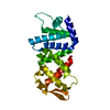
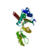
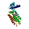
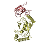
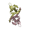
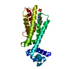
| Entry | Database: PDB / ID: 1xt0 | ||||||
|---|---|---|---|---|---|---|---|
| Title | The Structure of N-terminal Sec7 domain of RalF | ||||||
 Components Components | guanine nucleotide exchange protein | ||||||
 Keywords Keywords |  SIGNALING PROTEIN / The N-terminal Sec7 domain of RalF SIGNALING PROTEIN / The N-terminal Sec7 domain of RalF | ||||||
| Function / homology |  Function and homology information Function and homology informationregulation of ARF protein signal transduction / guanyl-nucleotide exchange factor activity Similarity search - Function | ||||||
| Biological species |   Legionella pneumophila (bacteria) Legionella pneumophila (bacteria) | ||||||
| Method | X-RAY DIFFRACTION / SYNCHROTRON / MOLECULAR REPLACEMENT / Resolution: 2.16 Å | ||||||
 Authors Authors | Amor, J.C. / Swails, J. / Roy, C.R. / Nagai, H. / Ingmundson, A. / Cheng, X. / Kahn, R.A. | ||||||
 Citation Citation | Journal: J.Biol.Chem. / Year: 2005 Title: The structure of RalF, an ADP-ribosylation factor guanine nucleotide exchange factor from Legionella pneumophila, reveals the presence of a cap over the active site Authors: Amor, J.C. / Swails, J. / Zhu, X. / Roy, C.R. / Nagai, H. / Ingmundson, A. / Cheng, X. / Kahn, R.A. | ||||||
| History |
|
- Structure visualization
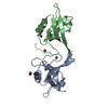
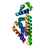
| Structure viewer | Molecule: MolmilJmol/JSmol |
|---|
- Downloads & links
-Download
| PDBx/mmCIF format | 1xt0.cif.gz | 54.4 KB | Display | PDBx/mmCIF format |
|---|---|---|---|---|
| PDB format | pdb1xt0.ent.gz | 38.6 KB | Display | PDB format |
| PDBx/mmJSON format | 1xt0.json.gz | Tree view | PDBx/mmJSON format | |
| Others |  Other downloads Other downloads |
-Validation report
| Arichive directory | https://data.pdbj.org/pub/pdb/validation_reports/xt/1xt0ftp://data.pdbj.org/pub/pdb/validation_reports/xt/1xt0 | HTTPS FTP |
|---|
-Related structure data
| Related structure data |  1xszSC S: Starting model for refinement C: citing same article ( |
|---|---|
| Similar structure data |

-Links
 PDBj
PDBj- Assembly
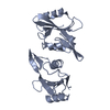
| Deposited unit | 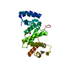
| ||||||||
|---|---|---|---|---|---|---|---|---|---|
| 1 |
| ||||||||
| Unit cell |
|
-Components
| #1: Protein | Mass: 23076.439 Da / Num. of mol.: 1 Source method: isolated from a genetically manipulated source Source: (gene. exp.) Legionella pneumophila (bacteria) / Plasmid: pHis-1 / Species (production host): Escherichia coli / Production host: Escherichia coli BL21(DE3) (bacteria) / Strain (production host): BL21(DE3) / References: UniProt: Q8RT31 |
|---|---|
| #2: Water | ChemComp-HOH / Water Mass: 18.015 Da / Num. of mol.: 73 / Source method: isolated from a natural source / Formula: H2O Mass: 18.015 Da / Num. of mol.: 73 / Source method: isolated from a natural source / Formula: H2O |
-Experimental details
-Experiment
| Experiment | Method: X-RAY DIFFRACTION / Number of used crystals: 1 |
|---|
- Sample preparation
| Crystal | Density Matthews: 2.06 Å3/Da / Density % sol: 40.15 % |
|---|---|
| Crystal grow | Temperature: 294 K / Method: vapor diffusion, sitting drop / pH: 8.5 Details: PEG2K, pH 8.5, VAPOR DIFFUSION, SITTING DROP, temperature 294K |
-Data collection
| Diffraction | Mean temperature: 108 K |
|---|---|
| Diffraction source | Source: SYNCHROTRON / Site: NSLS  / Beamline: X26C / Wavelength: 0.96 Å / Beamline: X26C / Wavelength: 0.96 Å |
| Detector | Type: ADSC QUANTUM 4 / Detector: CCD / Date: Aug 27, 2003 |
| Radiation | Protocol: MAD / Monochromatic (M) / Laue (L): M / Scattering type: x-ray |
| Radiation wavelength | Wavelength: 0.96 Å / Relative weight: 1 |
| Reflection | Resolution: 2.16→50 Å / Num. all: 17589 / Num. obs: 17589 / % possible obs: 91 % / Observed criterion σ(F): 0 / Observed criterion σ(I): -3 / Redundancy: 4.6 % / Biso Wilson estimate: 17.9 Å2 / Rmerge(I) obs: 0.066 / Net I/σ(I): 12.1 |
| Reflection shell | Resolution: 2.16→2.24 Å / Rmerge(I) obs: 0.115 / Mean I/σ(I) obs: 6.1 / Num. unique all: 1821 / % possible all: 92.6 |
- Processing
| Software |
| ||||||||||||||||||||||||||||||||||||
|---|---|---|---|---|---|---|---|---|---|---|---|---|---|---|---|---|---|---|---|---|---|---|---|---|---|---|---|---|---|---|---|---|---|---|---|---|---|
| Refinement | Method to determine structure: MOLECULAR REPLACEMENT Starting model: 1XSZ Resolution: 2.16→50 Å / Rfactor Rfree error: 0.007 / Data cutoff high absF: 582242.4 / Data cutoff low absF: 0 / Isotropic thermal model: Isotropic / Cross valid method: THROUGHOUT / σ(F): 0 / Stereochemistry target values: Engh & Huber
| ||||||||||||||||||||||||||||||||||||
| Solvent computation | Solvent model: FLAT MODEL / Bsol: 47.0814 Å2 / ksol: 0.381791 e/Å3 | ||||||||||||||||||||||||||||||||||||
| Displacement parameters | Biso mean: 30.2 Å2
| ||||||||||||||||||||||||||||||||||||
| Refine analyze |
| ||||||||||||||||||||||||||||||||||||
| Refinement step | Cycle: LAST / Resolution: 2.16→50 Å
| ||||||||||||||||||||||||||||||||||||
| Refine LS restraints |
| ||||||||||||||||||||||||||||||||||||
| LS refinement shell | Resolution: 2.16→2.24 Å / Rfactor Rfree error: 0.024 / Total num. of bins used: 10
| ||||||||||||||||||||||||||||||||||||
| Xplor file |
|
