 Movie
Movie Controller

[English] 日本語
 Yorodumi
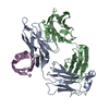
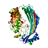
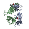
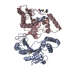
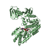
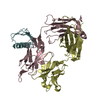
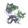
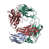
- PDB-2r6z: Crystal structure of the SAM-dependent methyltransferase NGO1261 ... -
+ Open data

- Basic information
| Entry | Database: PDB / ID: 2r6z | ||||||
|---|---|---|---|---|---|---|---|
| Title | Crystal structure of the SAM-dependent methyltransferase NGO1261 from Neisseria gonorrhoeae, Northeast Structural Genomics Consortium Target NgR48 | ||||||
 Components Components | UPF0341 protein in rsp 3' region | ||||||
 Keywords Keywords |  TRANSFERASE / alpha-beta protein / Structural Genomics / PSI-2 / Protein Structure Initiative / Northeast Structural Genomics Consortium / NESG TRANSFERASE / alpha-beta protein / Structural Genomics / PSI-2 / Protein Structure Initiative / Northeast Structural Genomics Consortium / NESG | ||||||
| Function / homology |  Function and homology information16S rRNA (guanine1516-N2)-methyltransferase / rRNA (guanine-N2-)-methyltransferase activity / cytoplasm Function and homology information16S rRNA (guanine1516-N2)-methyltransferase / rRNA (guanine-N2-)-methyltransferase activity / cytoplasmSimilarity search - Function | ||||||
| Biological species |  Neisseria gonorrhoeae (bacteria) Neisseria gonorrhoeae (bacteria) | ||||||
| Method | X-RAY DIFFRACTION / SYNCHROTRON / SAD / Resolution: 1.8 Å | ||||||
 Authors Authors | Forouhar, F. / Abashidze, M. / Seetharaman, J. / Mao, L. / Nwosu, C. / Fang, Y. / Xiao, R. / Baran, M.C. / Acton, T.B. / Montelione, G.T. ...Forouhar, F. / Abashidze, M. / Seetharaman, J. / Mao, L. / Nwosu, C. / Fang, Y. / Xiao, R. / Baran, M.C. / Acton, T.B. / Montelione, G.T. / Tong, L. / Hunt, J.F. / Northeast Structural Genomics Consortium (NESG) | ||||||
 Citation Citation | Journal: To be Published Title: Crystal structure of the SAM-dependent methyltransferase NGO1261 from Neisseria gonorrhoeae. Authors: Forouhar, F. / Abashidze, M. / Seetharaman, J. / Mao, L. / Nwosu, C. / Fang, Y. / Xiao, R. / Baran, M.C. / Acton, T.B. / Montelione, G.T. / Tong, L. / Hunt, J.F. / Northeast Structural Genomics Consortium (NESG) | ||||||
| History |
|
- Structure visualization
| Structure viewer | Molecule: MolmilJmol/JSmol |
|---|
- Downloads & links
-Download
| PDBx/mmCIF format | 2r6z.cif.gz | 107.6 KB | Display | PDBx/mmCIF format |
|---|---|---|---|---|
| PDB format | pdb2r6z.ent.gz | 87.4 KB | Display | PDB format |
| PDBx/mmJSON format | 2r6z.json.gz | Tree view | PDBx/mmJSON format | |
| Others |  Other downloads Other downloads |
-Validation report
| Arichive directory | https://data.pdbj.org/pub/pdb/validation_reports/r6/2r6zftp://data.pdbj.org/pub/pdb/validation_reports/r6/2r6z | HTTPS FTP |
|---|
-Related structure data
| Similar structure data | |
|---|---|
| Other databases |


-Links
 PDBj
PDBj- Assembly
| Deposited unit | 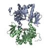
| ||||||||
|---|---|---|---|---|---|---|---|---|---|
| 1 |
| ||||||||
| Unit cell |
|
-Components
| #1: Protein | Mass: 28612.930 Da / Num. of mol.: 2 Source method: isolated from a genetically manipulated source Source: (gene. exp.) Neisseria gonorrhoeae (bacteria) / Strain: MS11, FA 1090 / Gene: NGO1261 / Plasmid: pET21 / Production host: Escherichia coli (E. coli) / Strain (production host): BL21(DE3)+Magic / References: UniProt: P72077#2: Water | ChemComp-HOH / | Water Mass: 18.015 Da / Num. of mol.: 453 / Source method: isolated from a natural source / Formula: H2O Mass: 18.015 Da / Num. of mol.: 453 / Source method: isolated from a natural source / Formula: H2O |
|---|
-Experimental details
-Experiment
| Experiment | Method: X-RAY DIFFRACTION / Number of used crystals: 1 |
|---|
- Sample preparation
| Crystal | Density Matthews: 2.26 Å3/Da / Density % sol: 45.48 % Description: The structure factor file contains Friedel pairs |
|---|---|
| Crystal grow | Temperature: 277 K / Method: microbatch under oil / pH: 6.5 Details: Protein solution: 10 mM Tris-HCl pH 7.5, 100 mM NaCl, 5 mM DTT. Reservoir solution: 100 mM Cacodylic acid pH 6.5, 18% PEG 3350, MICROBATCH UNDER OIL, temperature 277K |
-Data collection
| Diffraction | Mean temperature: 100 K |
|---|---|
| Diffraction source | Source: SYNCHROTRON / Site: NSLS  / Beamline: X4C / Wavelength: 0.97893 Å / Beamline: X4C / Wavelength: 0.97893 Å |
| Detector | Type: ADSC QUANTUM 210 / Detector: CCD / Date: Aug 29, 2007 / Details: mirrors |
| Radiation | Monochromator: Si 111 CHANNEL / Protocol: SINGLE WAVELENGTH / Monochromatic (M) / Laue (L): M / Scattering type: x-ray |
| Radiation wavelength | Wavelength: 0.97893 Å / Relative weight: 1 |
| Reflection | Resolution: 1.8→26.82 Å / Num. all: 91274 / Num. obs: 91274 / % possible obs: 98.4 % / Observed criterion σ(F): 0 / Observed criterion σ(I): 0 / Redundancy: 4.1 % / Biso Wilson estimate: 14.9 Å2 / Rmerge(I) obs: 0.04 / Rsym value: 0.031 / Net I/σ(I): 28.9 |
| Reflection shell | Resolution: 1.8→1.86 Å / Redundancy: 3.9 % / Rmerge(I) obs: 0.365 / Mean I/σ(I) obs: 2.96 / Num. unique all: 8942 / Rsym value: 0.27 / % possible all: 96.6 |
- Processing
| Software |
| |||||||||||||||||||||||||
|---|---|---|---|---|---|---|---|---|---|---|---|---|---|---|---|---|---|---|---|---|---|---|---|---|---|---|
| Refinement | Method to determine structure: SAD / Resolution: 1.8→26.82 Å / Rfactor Rfree error: 0.003 / Data cutoff high absF: 513500.77 / Data cutoff low absF: 0 / Isotropic thermal model: OVERALL / Cross valid method: THROUGHOUT / σ(F): 2 / Stereochemistry target values: Engh & Huber / Details: The Friedel pairs were used for phasing
| |||||||||||||||||||||||||
| Solvent computation | Solvent model: FLAT MODEL / Bsol: 58.7004 Å2 / ksol: 0.4 e/Å3 | |||||||||||||||||||||||||
| Displacement parameters | Biso mean: 28 Å2
| |||||||||||||||||||||||||
| Refine analyze |
| |||||||||||||||||||||||||
| Refinement step | Cycle: LAST / Resolution: 1.8→26.82 Å
| |||||||||||||||||||||||||
| Refine LS restraints |
| |||||||||||||||||||||||||
| LS refinement shell | Resolution: 1.8→1.86 Å / Rfactor Rfree error: 0.013 / Total num. of bins used: 10
|
