 Movie
Movie Controller


+ Open data

- Basic information
| Entry | Database: PDB / ID: 1mhh | ||||||
|---|---|---|---|---|---|---|---|
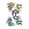
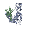
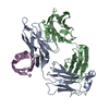
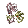
| Title | Structure of P. magnus protein L mutant bound to a mouse Fab | ||||||
 Components Components |
| ||||||
 Keywords Keywords |  IMMUNE SYSTEM / Antibody-antigen complex / B cell superantigen / Immunoglobulin binding protein IMMUNE SYSTEM / Antibody-antigen complex / B cell superantigen / Immunoglobulin binding protein | ||||||
| Function / homology | Ubiquitin-like (UB roll) - #10 / Ubiquitin-like (UB roll) / Immunoglobulins / Roll / Immunoglobulin-like / Sandwich / Mainly Beta / Alpha Beta Function and homology information Function and homology information | ||||||
| Biological species |  Finegoldia magna (bacteria) Finegoldia magna (bacteria) Mus musculus (house mouse) Mus musculus (house mouse) | ||||||
| Method | X-RAY DIFFRACTION / SYNCHROTRON / MOLECULAR REPLACEMENT / Resolution: 2.1 Å | ||||||
 Authors Authors | Graille, M. / Stura, E.A. | ||||||
 Citation Citation | Journal: J.Biol.Chem. / Year: 2002 Title: Evidence for plasticity and structural mimicry at the immunoglobulin light chain-protein L interface Authors: Graille, M. / Harrison, S. / Crump, M.P. / Findlow, S.C. / Housden, N.G. / Muller, B.H. / Battail-Poirot, N. / Sibai, G. / Sutton, B.J. / Taussig, M.J. / Jolivet-Reynaud, C. / Gore, M.G. / Stura, E.A. #1: Journal: Structure / Year: 2001Title: Complex between Peptostreptococcus Magnus Protein L and a Human Antibody Reveals Structural Convergence in the Interaction Modes of Fab Binding Modes Authors: Graille, M. / Stura, E.A. / Housden, N.G. / Beckingham, J.A. / Bottomley, S.P. / Beale, D. / Taussig, M.J. / Sutton, B.J. / Gore, M.G. / Charbonnier, J.-B. | ||||||
| History |
|
- Structure visualization
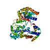
| Structure viewer | Molecule: MolmilJmol/JSmol |
|---|
- Downloads & links
-Download
| PDBx/mmCIF format | 1mhh.cif.gz | 216 KB | Display | PDBx/mmCIF format |
|---|---|---|---|---|
| PDB format | pdb1mhh.ent.gz | 178 KB | Display | PDB format |
| PDBx/mmJSON format | 1mhh.json.gz | Tree view | PDBx/mmJSON format | |
| Others |  Other downloads Other downloads |
-Validation report
| Arichive directory | https://data.pdbj.org/pub/pdb/validation_reports/mh/1mhhftp://data.pdbj.org/pub/pdb/validation_reports/mh/1mhh | HTTPS FTP |
|---|
-Related structure data
| Related structure data | |
|---|---|
| Similar structure data |


-Links
 PDBj
PDBj

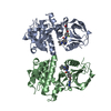
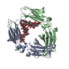
- Assembly
| Deposited unit | 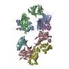
| ||||||||
|---|---|---|---|---|---|---|---|---|---|
| 1 | 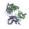
| ||||||||
| 2 | 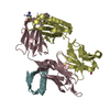
| ||||||||
| Unit cell |
|
-Components
| #1: Antibody | Fragment antigen-binding Mass: 24391.102 Da / Num. of mol.: 2 / Source method: isolated from a natural source / Source: (natural) Mus musculus (house mouse)#2: Antibody | Fragment antigen-bindingMass: 23406.236 Da / Num. of mol.: 2 / Source method: isolated from a natural source / Source: (natural) Mus musculus (house mouse)#3: Protein | Mass: 6915.743 Da / Num. of mol.: 2 / Mutation: D855A/Y864W Source method: isolated from a genetically manipulated source Source: (gene. exp.) Finegoldia magna (bacteria) / Strain: ATCC 29328 / Plasmid: pKK233-3 / Production host: Escherichia coli (E. coli) / Strain (production host): JM103#4: Chemical | Ethylene glycol  Mass: 62.068 Da / Num. of mol.: 2 / Source method: obtained synthetically / Formula: C2H6O2 Mass: 62.068 Da / Num. of mol.: 2 / Source method: obtained synthetically / Formula: C2H6O2#5: Water | ChemComp-HOH / | Water Mass: 18.015 Da / Num. of mol.: 739 / Source method: isolated from a natural source / Formula: H2O Mass: 18.015 Da / Num. of mol.: 739 / Source method: isolated from a natural source / Formula: H2O |
|---|
-Experimental details
-Experiment
| Experiment | Method: X-RAY DIFFRACTION / Number of used crystals: 2 |
|---|
- Sample preparation
| Crystal | Density Matthews: 2.7 Å3/Da / Density % sol: 54.42 % | ||||||||||||||||||||||||||||||
|---|---|---|---|---|---|---|---|---|---|---|---|---|---|---|---|---|---|---|---|---|---|---|---|---|---|---|---|---|---|---|---|
| Crystal grow | Temperature: 293 K / Method: vapor diffusion, sitting drop / pH: 4.5 Details: 10%(wt/wt) MPEG 5K, 100mM sodium acetate, pH 4.5, VAPOR DIFFUSION, SITTING DROP, temperature 293K | ||||||||||||||||||||||||||||||
| Crystal grow | *PLUS | ||||||||||||||||||||||||||||||
| Components of the solutions | *PLUS
|
-Data collection
| Diffraction |
| |||||||||||||||
|---|---|---|---|---|---|---|---|---|---|---|---|---|---|---|---|---|
| Diffraction source |
| |||||||||||||||
| Detector |
| |||||||||||||||
| Radiation | Protocol: SINGLE WAVELENGTH / Monochromatic (M) / Laue (L): M / Scattering type: x-ray | |||||||||||||||
| Radiation wavelength |
| |||||||||||||||
| Reflection | Resolution: 2.1→20 Å / Num. all: 69885 / Num. obs: 69723 / % possible obs: 97.6 % / Observed criterion σ(F): 2 / Observed criterion σ(I): 2 / Redundancy: 6.77 % / Biso Wilson estimate: 38 Å2 / Rsym value: 0.067 / Net I/σ(I): 26.6 | |||||||||||||||
| Reflection shell | Resolution: 2.1→2.16 Å / Redundancy: 5.86 % / Mean I/σ(I) obs: 3.5 / Num. unique all: 4986 / Rsym value: 0.417 / % possible all: 85 | |||||||||||||||
| Reflection | *PLUS Lowest resolution: 20 Å / Rmerge(I) obs: 0.067 | |||||||||||||||
| Reflection shell | *PLUS % possible obs: 85 % / Rmerge(I) obs: 0.417 |

- Processing
| Software |
| |||||||||||||||||||||||||
|---|---|---|---|---|---|---|---|---|---|---|---|---|---|---|---|---|---|---|---|---|---|---|---|---|---|---|
| Refinement | Method to determine structure: MOLECULAR REPLACEMENT / Resolution: 2.1→20 Å / Cross valid method: THROUGHOUT / σ(F): 2 / Stereochemistry target values: Engh & Huber
| |||||||||||||||||||||||||
| Displacement parameters |
| |||||||||||||||||||||||||
| Refine analyze | Luzzati coordinate error obs: 0.24 Å | |||||||||||||||||||||||||
| Refinement step | Cycle: LAST / Resolution: 2.1→20 Å
| |||||||||||||||||||||||||
| Refine LS restraints |
| |||||||||||||||||||||||||
| LS refinement shell | Resolution: 2.1→2.11 Å
| |||||||||||||||||||||||||
| Refinement | *PLUS Lowest resolution: 20 Å / σ(F): 2 / % reflection Rfree: 5 % | |||||||||||||||||||||||||
| Solvent computation | *PLUS | |||||||||||||||||||||||||
| Displacement parameters | *PLUS | |||||||||||||||||||||||||
| Refine LS restraints | *PLUS Type: c_angle_deg / Dev ideal: 1.3 | |||||||||||||||||||||||||
| LS refinement shell | *PLUS Highest resolution: 2.1 Å / Rfactor Rfree: 0.236 / Rfactor Rwork: 0.223 |
