 Movie
Movie Controller


+ Open data

- Basic information
| Entry | Database: PDB / ID: 1ymh | ||||||
|---|---|---|---|---|---|---|---|
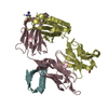
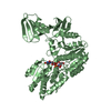
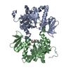
| Title | anti-HCV Fab 19D9D6 complexed with protein L (PpL) mutant A66W | ||||||
 Components Components |
| ||||||
 Keywords Keywords |  IMMUNE SYSTEM / Engineering of Crystal contacts / PpL-Fab complex IMMUNE SYSTEM / Engineering of Crystal contacts / PpL-Fab complex | ||||||
| Function / homology |  Function and homology information Function and homology information | ||||||
| Biological species |  Finegoldia magna (bacteria) Finegoldia magna (bacteria) Mus musculus (house mouse) Mus musculus (house mouse) | ||||||
| Method | X-RAY DIFFRACTION / SYNCHROTRON / MOLECULAR REPLACEMENT / Resolution: 2.6 Å | ||||||
 Authors Authors | Granata, V. / Housden, N.G. / Harrison, S. / Jolivet-Reynaud, C. / Gore, M.G. / Stura, E.A. | ||||||
 Citation Citation | Journal: Acta Crystallogr.,Sect.D / Year: 2005 Title: Comparison of the crystallization and crystal packing of two Fab single-site mutant protein L complexes. Authors: Granata, V. / Housden, N.G. / Harrison, S. / Jolivet-Reynaud, C. / Gore, M.G. / Stura, E.A. | ||||||
| History |
|
- Structure visualization
| Structure viewer | Molecule: MolmilJmol/JSmol |
|---|
- Downloads & links
-Download
| PDBx/mmCIF format | 1ymh.cif.gz | 219 KB | Display | PDBx/mmCIF format |
|---|---|---|---|---|
| PDB format | pdb1ymh.ent.gz | 166.7 KB | Display | PDB format |
| PDBx/mmJSON format | 1ymh.json.gz | Tree view | PDBx/mmJSON format | |
| Others |  Other downloads Other downloads |
-Validation report
| Arichive directory | https://data.pdbj.org/pub/pdb/validation_reports/ym/1ymhftp://data.pdbj.org/pub/pdb/validation_reports/ym/1ymh | HTTPS FTP |
|---|
-Related structure data
| Related structure data | 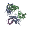 1mhhS S: Starting model for refinement |
|---|---|
| Similar structure data |


-Links
 PDBj
PDBj

- Assembly
| Deposited unit | 
| ||||||||
|---|---|---|---|---|---|---|---|---|---|
| 1 | 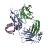
| ||||||||
| 2 | 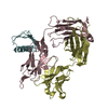
| ||||||||
| Unit cell |
|
-Components
| #1: Antibody | Mass: 24334.051 Da / Num. of mol.: 2 / Source method: isolated from a natural source / Source: (natural) Mus musculus (house mouse) / Cell line: Hybridoma#2: Antibody | Mass: 23563.430 Da / Num. of mol.: 2 / Source method: isolated from a natural source / Source: (natural) Mus musculus (house mouse) / Cell line: Hybridoma#3: Protein | / PpLMass: 7310.145 Da / Num. of mol.: 2 / Mutation: A866W Source method: isolated from a genetically manipulated source Source: (gene. exp.) Finegoldia magna (bacteria) / Strain: ATCC 29328 / Plasmid: PKK223-3 / Production host: Escherichia coli (E. coli) / References: UniProt: Q51918#4: Water | ChemComp-HOH / | Water Mass: 18.015 Da / Num. of mol.: 116 / Source method: isolated from a natural source / Formula: H2O Mass: 18.015 Da / Num. of mol.: 116 / Source method: isolated from a natural source / Formula: H2O |
|---|
-Experimental details
-Experiment
| Experiment | Method: X-RAY DIFFRACTION / Number of used crystals: 1 |
|---|
- Sample preparation
| Crystal | Density Matthews: 2.5 Å3/Da / Density % sol: 51.1 % |
|---|---|
| Crystal grow | Temperature: 293 K / Method: vapor diffusion, sitting drop / pH: 5.1 Details: 9% PEG5K, 100mM Na cacodylate, pH 5.1, VAPOR DIFFUSION, SITTING DROP, temperature 293K |
-Data collection
| Diffraction | Mean temperature: 100 K |
|---|---|
| Diffraction source | Source: SYNCHROTRON / Site: ESRF  / Beamline: ID29 / Wavelength: 0.720169 Å / Beamline: ID29 / Wavelength: 0.720169 Å |
| Detector | Type: ADSC QUANTUM 210 / Detector: CCD / Date: Feb 5, 2004 |
| Radiation | Monochromator: Si / Protocol: SINGLE WAVELENGTH / Monochromatic (M) / Laue (L): M / Scattering type: x-ray |
| Radiation wavelength | Wavelength: 0.720169 Å / Relative weight: 1 |
| Reflection | Resolution: 2.6→88 Å / Num. all: 39989 / Num. obs: 39109 / % possible obs: 97.8 % / Observed criterion σ(F): 0 / Observed criterion σ(I): 0 / Redundancy: 11 % / Biso Wilson estimate: 79.237 Å2 / Rmerge(I) obs: 0.058 / Net I/σ(I): 15 |
| Reflection shell | Resolution: 2.6→2.66 Å / Redundancy: 11 % / Rmerge(I) obs: 0.489 / Mean I/σ(I) obs: 4 / Num. unique all: 2806 / % possible all: 100 |
- Processing
| Software |
| ||||||||||||||||||||||||||||||||||||||||||||||||||||||||||||||||||||||||||||||||||||||||||||||||||||||||||||||||||||||||||||||||||||||||||||||||||||||||||||||||||||||||||
|---|---|---|---|---|---|---|---|---|---|---|---|---|---|---|---|---|---|---|---|---|---|---|---|---|---|---|---|---|---|---|---|---|---|---|---|---|---|---|---|---|---|---|---|---|---|---|---|---|---|---|---|---|---|---|---|---|---|---|---|---|---|---|---|---|---|---|---|---|---|---|---|---|---|---|---|---|---|---|---|---|---|---|---|---|---|---|---|---|---|---|---|---|---|---|---|---|---|---|---|---|---|---|---|---|---|---|---|---|---|---|---|---|---|---|---|---|---|---|---|---|---|---|---|---|---|---|---|---|---|---|---|---|---|---|---|---|---|---|---|---|---|---|---|---|---|---|---|---|---|---|---|---|---|---|---|---|---|---|---|---|---|---|---|---|---|---|---|---|---|---|---|
| Refinement | Method to determine structure: MOLECULAR REPLACEMENT Starting model: PDB ENTRY 1MHH Resolution: 2.6→87.71 Å / Cor.coef. Fo:Fc: 0.939 / Cor.coef. Fo:Fc free: 0.893 / SU B: 13.137 / SU ML: 0.283 / Cross valid method: THROUGHOUT / σ(F): 0 / σ(I): 0 / ESU R: 0.71 / ESU R Free: 0.372 / Stereochemistry target values: MAXIMUM LIKELIHOOD / Details: HYDROGENS HAVE BEEN ADDED IN THE RIDING POSITIONS
| ||||||||||||||||||||||||||||||||||||||||||||||||||||||||||||||||||||||||||||||||||||||||||||||||||||||||||||||||||||||||||||||||||||||||||||||||||||||||||||||||||||||||||
| Solvent computation | Ion probe radii: 0.8 Å / Shrinkage radii: 0.8 Å / VDW probe radii: 1.4 Å / Solvent model: BABINET MODEL WITH MASK | ||||||||||||||||||||||||||||||||||||||||||||||||||||||||||||||||||||||||||||||||||||||||||||||||||||||||||||||||||||||||||||||||||||||||||||||||||||||||||||||||||||||||||
| Displacement parameters | Biso mean: 64.086 Å2
| ||||||||||||||||||||||||||||||||||||||||||||||||||||||||||||||||||||||||||||||||||||||||||||||||||||||||||||||||||||||||||||||||||||||||||||||||||||||||||||||||||||||||||
| Refinement step | Cycle: LAST / Resolution: 2.6→87.71 Å
| ||||||||||||||||||||||||||||||||||||||||||||||||||||||||||||||||||||||||||||||||||||||||||||||||||||||||||||||||||||||||||||||||||||||||||||||||||||||||||||||||||||||||||
| Refine LS restraints |
| ||||||||||||||||||||||||||||||||||||||||||||||||||||||||||||||||||||||||||||||||||||||||||||||||||||||||||||||||||||||||||||||||||||||||||||||||||||||||||||||||||||||||||
| LS refinement shell | Resolution: 2.603→2.671 Å / Total num. of bins used: 20 /
|
