 Movie
Movie Controller


+ Open data

- Basic information
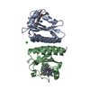
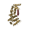
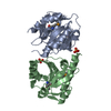
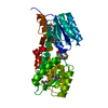
| Entry | Database: PDB / ID: 2qvl | ||||||
|---|---|---|---|---|---|---|---|
| Title | Crystal Structure of Diacylglycerol Kinase | ||||||
 Components Components | Diacylglycerol Kinase DgkB | ||||||
 Keywords Keywords |  TRANSFERASE / alpha-beta domain 1 / beta sandwich domain 2 / Native protein TRANSFERASE / alpha-beta domain 1 / beta sandwich domain 2 / Native protein | ||||||
| Function / homology |  Function and homology information Function and homology informationdiacylglycerol kinase (ATP) / ATP-dependent diacylglycerol kinase activity / phospholipid biosynthetic process / phosphorylation / ATP binding / identical protein binding / metal ion bindingSimilarity search - Function | ||||||
| Biological species |   Staphylococcus aureus (bacteria) Staphylococcus aureus (bacteria) | ||||||
| Method | X-RAY DIFFRACTION / SYNCHROTRON / SAD / Resolution: 2.4 Å | ||||||
 Authors Authors | Miller, D.J. / Jerga, A. / Rock, C.O. / White, S.W. | ||||||
 Citation Citation | Journal: Structure / Year: 2008 Title: Analysis of the Staphylococcus aureus DgkB Structure Reveals a Common Catalytic Mechanism for the Soluble Diacylglycerol Kinases. Authors: Miller, D.J. / Jerga, A. / Rock, C.O. / White, S.W. | ||||||
| History |
|
- Structure visualization
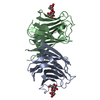
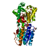
| Structure viewer | Molecule: MolmilJmol/JSmol |
|---|
- Downloads & links
-Download
| PDBx/mmCIF format | 2qvl.cif.gz | 66.9 KB | Display | PDBx/mmCIF format |
|---|---|---|---|---|
| PDB format | pdb2qvl.ent.gz | 52.6 KB | Display | PDB format |
| PDBx/mmJSON format | 2qvl.json.gz | Tree view | PDBx/mmJSON format | |
| Others |  Other downloads Other downloads |
-Validation report
| Arichive directory | https://data.pdbj.org/pub/pdb/validation_reports/qv/2qvlftp://data.pdbj.org/pub/pdb/validation_reports/qv/2qvl | HTTPS FTP |
|---|
-Related structure data


-Links
 PDBj
PDBj- Assembly
| Deposited unit | 
| ||||||||
|---|---|---|---|---|---|---|---|---|---|
| 1 |
| ||||||||
| 2 | 
| ||||||||
| Unit cell |
|
-Components
| #1: Protein | Mass: 37899.238 Da / Num. of mol.: 1 Source method: isolated from a genetically manipulated source Source: (gene. exp.) Staphylococcus aureus (bacteria) / Gene: SAR1989 / Plasmid: pAJ015 / Production host: Escherichia coli (E. coli) / Strain (production host): methionine auxotroph B834 / References: UniProt: Q6GFF9, diacylglycerol kinase (ATP) |
|---|---|
| #2: Water | ChemComp-HOH / Water Mass: 18.015 Da / Num. of mol.: 116 / Source method: isolated from a natural source / Formula: H2O Mass: 18.015 Da / Num. of mol.: 116 / Source method: isolated from a natural source / Formula: H2O |
-Experimental details
-Experiment
| Experiment | Method: X-RAY DIFFRACTION / Number of used crystals: 1 |
|---|
- Sample preparation
| Crystal | Density Matthews: 2.47 Å3/Da / Density % sol: 50.16 % |
|---|---|
| Crystal grow | Temperature: 277 K / Method: vapor diffusion, sitting drop / pH: 8.5 Details: drop: 0.1 M Tris pH 8.5, 9% PEG2KMME; well: 0.1 M Tris pH 8.5, 18% PEG2KMME, VAPOR DIFFUSION, SITTING DROP, temperature 277K |
-Data collection
| Diffraction | Mean temperature: 170 K |
|---|---|
| Diffraction source | Source: SYNCHROTRON / Site: APS  / Beamline: 22-ID / Wavelength: 0.9794 Å / Beamline: 22-ID / Wavelength: 0.9794 Å |
| Detector | Type: MARMOSAIC 300 mm CCD / Detector: CCD / Date: Feb 9, 2007 / Details: mirrors |
| Radiation | Monochromator: Si-220 / Protocol: SINGLE WAVELENGTH / Monochromatic (M) / Laue (L): M / Scattering type: x-ray |
| Radiation wavelength | Wavelength: 0.9794 Å / Relative weight: 1 |
| Reflection | Resolution: 2.4→26.15 Å / Num. all: 15527 / Num. obs: 15472 / % possible obs: 99.8 % / Observed criterion σ(F): 0 / Observed criterion σ(I): 0 / Redundancy: 8.8 % / Rsym value: 0.094 / Net I/σ(I): 28.1 |
| Reflection shell | Resolution: 2.4→2.49 Å / Redundancy: 6.1 % / Rmerge(I) obs: 0.376 / Mean I/σ(I) obs: 8.2 / Num. unique all: 1491 / % possible all: 99.5 |
- Processing
| Software |
| ||||||||||||||||||||||||||||||||||||||||||||||||||||||||||||||||||||||||||||||||||||||||||
|---|---|---|---|---|---|---|---|---|---|---|---|---|---|---|---|---|---|---|---|---|---|---|---|---|---|---|---|---|---|---|---|---|---|---|---|---|---|---|---|---|---|---|---|---|---|---|---|---|---|---|---|---|---|---|---|---|---|---|---|---|---|---|---|---|---|---|---|---|---|---|---|---|---|---|---|---|---|---|---|---|---|---|---|---|---|---|---|---|---|---|---|
| Refinement | Method to determine structure: SAD / Resolution: 2.4→26.15 Å / Cor.coef. Fo:Fc: 0.923 / Cor.coef. Fo:Fc free: 0.913 / SU B: 8.462 / SU ML: 0.2 / Cross valid method: THROUGHOUT / σ(F): 0 / σ(I): 0 / ESU R: 0.389 / ESU R Free: 0.272 / Stereochemistry target values: MAXIMUM LIKELIHOOD / Details: HYDROGENS HAVE BEEN ADDED IN THE RIDING POSITIONS
| ||||||||||||||||||||||||||||||||||||||||||||||||||||||||||||||||||||||||||||||||||||||||||
| Solvent computation | Ion probe radii: 0.8 Å / Shrinkage radii: 0.8 Å / VDW probe radii: 1.2 Å / Solvent model: MASK | ||||||||||||||||||||||||||||||||||||||||||||||||||||||||||||||||||||||||||||||||||||||||||
| Displacement parameters | Biso mean: 39.969 Å2
| ||||||||||||||||||||||||||||||||||||||||||||||||||||||||||||||||||||||||||||||||||||||||||
| Refinement step | Cycle: LAST / Resolution: 2.4→26.15 Å
| ||||||||||||||||||||||||||||||||||||||||||||||||||||||||||||||||||||||||||||||||||||||||||
| Refine LS restraints |
| ||||||||||||||||||||||||||||||||||||||||||||||||||||||||||||||||||||||||||||||||||||||||||
| LS refinement shell | Resolution: 2.4→2.45 Å / Total num. of bins used: 20
|
