 Movie
Movie Controller


+ Open data

- Basic information
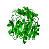
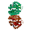
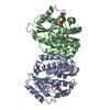
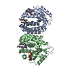
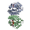
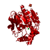
| Entry | Database: PDB / ID: 2qrn | ||||||
|---|---|---|---|---|---|---|---|
| Title | Human Deoxycytidine kinase dCMP, UDP, Mg ion product complex | ||||||
 Components Components | Deoxycytidine kinase | ||||||
 Keywords Keywords | TRANSFERASE / deoxycytidine kinase / deoxycytidine monophosphate / uridine diphosphate / ATP-binding / Nucleotide-binding / Nucleus / Phosphorylation | ||||||
| Function / homology |  Function and homology informationdeoxycytidine kinase / 2'-deoxyadenosine kinase / deoxyguanosine kinase / dAMP salvage / deoxycytidine kinase activity / nucleoside phosphate biosynthetic process / deoxyguanosine kinase activity / deoxyadenosine kinase activity / Pyrimidine salvage / cytidine kinase activity ...deoxycytidine kinase / 2'-deoxyadenosine kinase / deoxyguanosine kinase / dAMP salvage / deoxycytidine kinase activity / nucleoside phosphate biosynthetic process / deoxyguanosine kinase activity / deoxyadenosine kinase activity / Pyrimidine salvage / cytidine kinase activity / pyrimidine nucleotide metabolic process / Purine salvage / phosphorylation / protein homodimerization activity / mitochondrion / nucleoplasm / ATP binding / cytosol / cytoplasm Function and homology informationdeoxycytidine kinase / 2'-deoxyadenosine kinase / deoxyguanosine kinase / dAMP salvage / deoxycytidine kinase activity / nucleoside phosphate biosynthetic process / deoxyguanosine kinase activity / deoxyadenosine kinase activity / Pyrimidine salvage / cytidine kinase activity ...deoxycytidine kinase / 2'-deoxyadenosine kinase / deoxyguanosine kinase / dAMP salvage / deoxycytidine kinase activity / nucleoside phosphate biosynthetic process / deoxyguanosine kinase activity / deoxyadenosine kinase activity / Pyrimidine salvage / cytidine kinase activity / pyrimidine nucleotide metabolic process / Purine salvage / phosphorylation / protein homodimerization activity / mitochondrion / nucleoplasm / ATP binding / cytosol / cytoplasmSimilarity search - Function | ||||||
| Biological species |  Homo sapiens (human) Homo sapiens (human) | ||||||
| Method | X-RAY DIFFRACTION / SYNCHROTRON / MOLECULAR REPLACEMENT / Resolution: 3.4 Å | ||||||
 Authors Authors | Ealick, S.E. / Soriano, E.V. | ||||||
 Citation Citation | Journal: Acta Crystallogr.,Sect.D / Year: 2007 Title: Structures of human deoxycytidine kinase product complexes. Authors: Soriano, E.V. / Clark, V.C. / Ealick, S.E. | ||||||
| History |
|
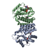
- Structure visualization
| Structure viewer | Molecule: MolmilJmol/JSmol |
|---|
- Downloads & links
-Download
| PDBx/mmCIF format | 2qrn.cif.gz | 187.8 KB | Display | PDBx/mmCIF format |
|---|---|---|---|---|
| PDB format | pdb2qrn.ent.gz | 151.4 KB | Display | PDB format |
| PDBx/mmJSON format | 2qrn.json.gz | Tree view | PDBx/mmJSON format | |
| Others |  Other downloads Other downloads |
-Validation report
| Arichive directory | https://data.pdbj.org/pub/pdb/validation_reports/qr/2qrnftp://data.pdbj.org/pub/pdb/validation_reports/qr/2qrn | HTTPS FTP |
|---|
-Related structure data
| Related structure data | 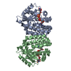 2qroC 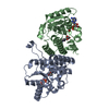 1p60S C: citing same article ( S: Starting model for refinement |
|---|---|
| Similar structure data |
-Links
 PDBj
PDBj


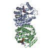
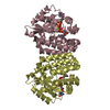
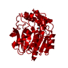
- Assembly
| Deposited unit | 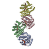
| ||||||||
|---|---|---|---|---|---|---|---|---|---|
| 1 | 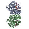
| ||||||||
| 2 | 
| ||||||||
| Unit cell |
|
-Components
| #1: Protein | / dCK Mass: 32723.850 Da / Num. of mol.: 4 Source method: isolated from a genetically manipulated source Source: (gene. exp.) Homo sapiens (human) / Gene: DCK / Production host:  Escherichia coli (E. coli) / References: UniProt: P27707, deoxycytidine kinase Escherichia coli (E. coli) / References: UniProt: P27707, deoxycytidine kinase#2: Chemical | ChemComp-MG /   Mass: 24.305 Da / Num. of mol.: 4 / Source method: obtained synthetically / Formula: Mg Mass: 24.305 Da / Num. of mol.: 4 / Source method: obtained synthetically / Formula: Mg#3: Chemical | ChemComp-UDP / Uridine diphosphate  Type: RNA linking / Mass: 404.161 Da / Num. of mol.: 4 / Source method: obtained synthetically / Formula: C9H14N2O12P2 / Comment: UDP*YM Type: RNA linking / Mass: 404.161 Da / Num. of mol.: 4 / Source method: obtained synthetically / Formula: C9H14N2O12P2 / Comment: UDP*YM#4: Chemical | ChemComp-DCM / Deoxycytidine monophosphate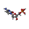  Mass: 307.197 Da / Num. of mol.: 4 / Source method: obtained synthetically / Formula: C9H14N3O7P Mass: 307.197 Da / Num. of mol.: 4 / Source method: obtained synthetically / Formula: C9H14N3O7P |
|---|
-Experimental details
-Experiment
| Experiment | Method: X-RAY DIFFRACTION / Number of used crystals: 1 |
|---|
- Sample preparation
| Crystal | Density Matthews: 2.86 Å3/Da / Density % sol: 57.05 % |
|---|---|
| Crystal grow | Temperature: 295 K / Method: vapor diffusion, hanging drop / pH: 7 Details: 16-17% ethanol,0.1 M HEPES, pH 7.0, VAPOR DIFFUSION, HANGING DROP, temperature 295K |
-Data collection
| Diffraction | Mean temperature: 100 K |
|---|---|
| Diffraction source | Source: SYNCHROTRON / Site: CHESS  / Beamline: A1 / Wavelength: 0.9764 Å / Beamline: A1 / Wavelength: 0.9764 Å |
| Detector | Type: ADSC QUANTUM 210 / Detector: CCD / Date: Jul 28, 2005 |
| Radiation | Protocol: SINGLE WAVELENGTH / Monochromatic (M) / Laue (L): M / Scattering type: x-ray |
| Radiation wavelength | Wavelength: 0.9764 Å / Relative weight: 1 |
| Reflection | Resolution: 3.4→48.12 Å / Num. all: 21038 / Num. obs: 19703 / % possible obs: 95.4 % / Observed criterion σ(F): 0 / Observed criterion σ(I): 0 / Redundancy: 5.4 % / Rmerge(I) obs: 0.129 / Rsym value: 0.129 / Net I/σ(I): 10.2 |
| Reflection shell | Resolution: 3.4→3.61 Å / Redundancy: 3.2 % / Mean I/σ(I) obs: 2.5 / Rsym value: 0.389 / % possible all: 73.3 |
- Processing
| Software |
| ||||||||||||||||||||||||||||||||||||
|---|---|---|---|---|---|---|---|---|---|---|---|---|---|---|---|---|---|---|---|---|---|---|---|---|---|---|---|---|---|---|---|---|---|---|---|---|---|
| Refinement | Method to determine structure: MOLECULAR REPLACEMENT Starting model: 1P60.pdb Resolution: 3.4→48.12 Å / Rfactor Rfree error: 0.009 / Data cutoff high absF: 158932.41 / Data cutoff low absF: 0 / Isotropic thermal model: RESTRAINED / Cross valid method: THROUGHOUT / σ(F): 0 / σ(I): 0 / Stereochemistry target values: Engh & Huber / Details: BULK SOLVENT MODEL USED
| ||||||||||||||||||||||||||||||||||||
| Solvent computation | Solvent model: FLAT MODEL / Bsol: 25.5115 Å2 / ksol: 0.3 e/Å3 | ||||||||||||||||||||||||||||||||||||
| Displacement parameters | Biso mean: 70.6 Å2
| ||||||||||||||||||||||||||||||||||||
| Refine analyze |
| ||||||||||||||||||||||||||||||||||||
| Refinement step | Cycle: LAST / Resolution: 3.4→48.12 Å
| ||||||||||||||||||||||||||||||||||||
| Refine LS restraints |
| ||||||||||||||||||||||||||||||||||||
| LS refinement shell | Resolution: 3.4→3.61 Å / Rfactor Rfree error: 0.036 / Total num. of bins used: 6
| ||||||||||||||||||||||||||||||||||||
| Xplor file |
|
