 Movie
Movie Controller

[English] 日本語
 Yorodumi
- PDB-2d0d: Crystal Structure of a Meta-cleavage Product Hydrolase (CumD) A12... -
+ Open data

- Basic information
| Entry | Database: PDB / ID: 2d0d | ||||||
|---|---|---|---|---|---|---|---|
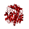
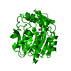
| Title | Crystal Structure of a Meta-cleavage Product Hydrolase (CumD) A129V Mutant | ||||||
 Components Components | 2-hydroxy-6-oxo-7-methylocta-2,4-dienoate hydrolase | ||||||
 Keywords Keywords |  HYDROLASE / alpha/beta-hydrolase / beta-ketolase / meta-cleavage product hydrolase / substrate specificity / cumene degradation / polychlorinated biphenyl degradation / PCB HYDROLASE / alpha/beta-hydrolase / beta-ketolase / meta-cleavage product hydrolase / substrate specificity / cumene degradation / polychlorinated biphenyl degradation / PCB | ||||||
| Function / homology |  Function and homology information Function and homology information | ||||||
| Biological species |  Pseudomonas fluorescens (bacteria) Pseudomonas fluorescens (bacteria) | ||||||
| Method | X-RAY DIFFRACTION / SYNCHROTRON / FOURIER SYNTHESIS / Resolution: 1.65 Å | ||||||
 Authors Authors | Jun, S.Y. / Fushinobu, S. / Nojiri, H. / Omori, T. / Shoun, H. / Wakagi, T. | ||||||
 Citation Citation | Journal: Biochim.Biophys.Acta / Year: 2006 Title: Improving the catalytic efficiency of a meta-cleavage product hydrolase (CumD) from Pseudomonas fluorescens IP01 Authors: Jun, S.Y. / Fushinobu, S. / Nojiri, H. / Omori, T. / Shoun, H. / Wakagi, T. #1: Journal: Biosci.Biotechnol.Biochem. / Year: 2005Title: A series of crystal structures of a meta-cleavage product hydrolase from Pseudomonas fluorescens IP01 (CumD) complexed with various cleavage products Authors: Fushinobu, S. / Jun, S.Y. / Hidaka, M. / Nojiri, H. / Yamane, H. / Shoun, H. / Omori, T. / Wakagi, T. #2: Journal: Protein Sci. / Year: 2002Title: Crystal structures of a meta-cleavage product hydrolase from Pseudomonas fluorescens IP01 (CumD) complexed with cleavage products Authors: Fushinobu, S. / Saku, T. / Hidaka, M. / Jun, S.Y. / Nojiri, H. / Yamane, H. / Shoun, H. / Omori, T. / Wakagi, T. | ||||||
| History |
|
- Structure visualization
| Structure viewer | Molecule: MolmilJmol/JSmol |
|---|
- Downloads & links
-Download
| PDBx/mmCIF format | 2d0d.cif.gz | 76.6 KB | Display | PDBx/mmCIF format |
|---|---|---|---|---|
| PDB format | pdb2d0d.ent.gz | 55.8 KB | Display | PDB format |
| PDBx/mmJSON format | 2d0d.json.gz | Tree view | PDBx/mmJSON format | |
| Others |  Other downloads Other downloads |
-Validation report
| Arichive directory | https://data.pdbj.org/pub/pdb/validation_reports/d0/2d0dftp://data.pdbj.org/pub/pdb/validation_reports/d0/2d0d | HTTPS FTP |
|---|
-Related structure data
| Related structure data |  1iupS S: Starting model for refinement |
|---|---|
| Similar structure data |


-Links
 PDBj
PDBj


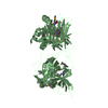
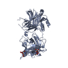
- Assembly
| Deposited unit | 
| ||||||||
|---|---|---|---|---|---|---|---|---|---|
| 1 | 
| ||||||||
| Unit cell |
| ||||||||
| Details | The second part of the biological assembly is generated by the two fold axis: x, -y+1, -z. |
-Components
| #1: Protein | Mass: 31550.658 Da / Num. of mol.: 1 / Mutation: A129V Source method: isolated from a genetically manipulated source Source: (gene. exp.) Pseudomonas fluorescens (bacteria) / Gene: CUMD / Plasmid: pIP140 / Production host: Escherichia coli (E. coli) / Strain (production host): JM109 / References: UniProt: P96965, EC: 3.7.1.9 | ||
|---|---|---|---|
| #2: Chemical | ChemComp-CL / Chloride  Mass: 35.453 Da / Num. of mol.: 1 / Source method: obtained synthetically / Formula: Cl Mass: 35.453 Da / Num. of mol.: 1 / Source method: obtained synthetically / Formula: Cl | ||
| #3: Chemical | Phosphate  Mass: 94.971 Da / Num. of mol.: 2 / Source method: obtained synthetically / Formula: PO4 Mass: 94.971 Da / Num. of mol.: 2 / Source method: obtained synthetically / Formula: PO4#4: Water | ChemComp-HOH / | Water Mass: 18.015 Da / Num. of mol.: 372 / Source method: isolated from a natural source / Formula: H2O Mass: 18.015 Da / Num. of mol.: 372 / Source method: isolated from a natural source / Formula: H2O |
-Experimental details
-Experiment
| Experiment | Method: X-RAY DIFFRACTION / Number of used crystals: 1 |
|---|
- Sample preparation
| Crystal | Density Matthews: 2.53 Å3/Da / Density % sol: 51 % |
|---|---|
| Crystal grow | Temperature: 298 K / Method: vapor diffusion, sitting drop / pH: 4.2 Details: PEG 3000, sodium phosphate/citrate, sodium chloride, pH 4.2, VAPOR DIFFUSION, SITTING DROP, temperature 298K |
-Data collection
| Diffraction | Mean temperature: 100 K |
|---|---|
| Diffraction source | Source: SYNCHROTRON / Site: Photon Factory  / Beamline: BL-6A / Wavelength: 1 Å / Beamline: BL-6A / Wavelength: 1 Å |
| Detector | Type: ADSC QUANTUM 4 / Detector: CCD / Date: Oct 22, 2004 |
| Radiation | Monochromator: Si 1 1 1 / Protocol: SINGLE WAVELENGTH / Monochromatic (M) / Laue (L): M / Scattering type: x-ray |
| Radiation wavelength | Wavelength: 1 Å / Relative weight: 1 |
| Reflection | Resolution: 1.65→46.84 Å / Num. all: 42760 / Num. obs: 42732 / % possible obs: 100 % / Observed criterion σ(F): 0 / Observed criterion σ(I): 0 / Biso Wilson estimate: 14.8 Å2 / Rsym value: 0.062 / Net I/σ(I): 26 |
| Reflection shell | Resolution: 1.65→1.71 Å / Mean I/σ(I) obs: 5.5 / Num. unique all: 4242 / Rsym value: 0.237 / % possible all: 100 |
- Processing
| Software |
| ||||||||||||||||||||||||||||||||||||||||||||||||||||||||||||||||||||||||||||||||
|---|---|---|---|---|---|---|---|---|---|---|---|---|---|---|---|---|---|---|---|---|---|---|---|---|---|---|---|---|---|---|---|---|---|---|---|---|---|---|---|---|---|---|---|---|---|---|---|---|---|---|---|---|---|---|---|---|---|---|---|---|---|---|---|---|---|---|---|---|---|---|---|---|---|---|---|---|---|---|---|---|---|
| Refinement | Method to determine structure: FOURIER SYNTHESIS Starting model: PDB ENTRY 1IUP Resolution: 1.65→46.84 Å / Rfactor Rfree error: 0.004 / Data cutoff high absF: 1954847.71 / Data cutoff low absF: 0 / Isotropic thermal model: RESTRAINED / Cross valid method: THROUGHOUT / σ(F): 0 / Stereochemistry target values: Engh & Huber
| ||||||||||||||||||||||||||||||||||||||||||||||||||||||||||||||||||||||||||||||||
| Solvent computation | Solvent model: FLAT MODEL / Bsol: 43.0574 Å2 / ksol: 0.343085 e/Å3 | ||||||||||||||||||||||||||||||||||||||||||||||||||||||||||||||||||||||||||||||||
| Displacement parameters | Biso mean: 14.7 Å2
| ||||||||||||||||||||||||||||||||||||||||||||||||||||||||||||||||||||||||||||||||
| Refine analyze |
| ||||||||||||||||||||||||||||||||||||||||||||||||||||||||||||||||||||||||||||||||
| Refinement step | Cycle: LAST / Resolution: 1.65→46.84 Å
| ||||||||||||||||||||||||||||||||||||||||||||||||||||||||||||||||||||||||||||||||
| Refine LS restraints |
| ||||||||||||||||||||||||||||||||||||||||||||||||||||||||||||||||||||||||||||||||
| LS refinement shell | Resolution: 1.65→1.75 Å / Rfactor Rfree error: 0.013 / Total num. of bins used: 6
| ||||||||||||||||||||||||||||||||||||||||||||||||||||||||||||||||||||||||||||||||
| Xplor file |
|
