 Movie
Movie Controller

[English] 日本語
 Yorodumi
- PDB-2qos: Crystal structure of complement protein C8 in complex with a pept... -
+ Open data

- Basic information
| Entry | Database: PDB / ID: 2qos | ||||||
|---|---|---|---|---|---|---|---|
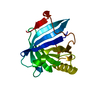
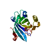
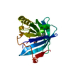
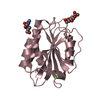
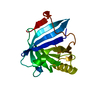
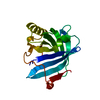
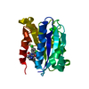
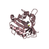
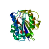
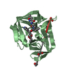
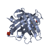
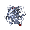
| Title | Crystal structure of complement protein C8 in complex with a peptide containing the C8 binding site on C8 | ||||||
 Components Components |
| ||||||
 Keywords Keywords |  IMMUNE SYSTEM / Beta barrel / lipocalin / Cleavage on pair of basic residues / Complement alternate pathway / Complement pathway / Cytolysis / EGF-like domain / Glycoprotein / Immune response / Innate immunity / Membrane attack complex / Polymorphism / Secreted IMMUNE SYSTEM / Beta barrel / lipocalin / Cleavage on pair of basic residues / Complement alternate pathway / Complement pathway / Cytolysis / EGF-like domain / Glycoprotein / Immune response / Innate immunity / Membrane attack complex / Polymorphism / Secreted | ||||||
| Function / homology |  Function and homology information Function and homology informationTerminal pathway of complement / membrane attack complex / complement binding / complement activation, alternative pathway / complement activation / retinol binding / complement activation, classical pathway / Regulation of Complement cascade / positive regulation of immune response / blood microparticle ...Terminal pathway of complement / membrane attack complex / complement binding / complement activation, alternative pathway / complement activation / retinol binding / complement activation, classical pathway / Regulation of Complement cascade / positive regulation of immune response / blood microparticle / killing of cells of another organism / immune response / protein-containing complex binding / extracellular space / extracellular exosome / extracellular region / membrane / plasma membraneSimilarity search - Function | ||||||
| Biological species |  Homo sapiens (human) Homo sapiens (human) | ||||||
| Method | X-RAY DIFFRACTION / SYNCHROTRON / Resolution: 1.81 Å | ||||||
 Authors Authors | Lovelace, L.L. / Chiswell, B. / Slade, D.J. / Sodetz, J.M. / Lebioda, L. | ||||||
 Citation Citation | Journal: Mol.Immunol. / Year: 2008 Title: Crystal structure of complement protein C8gamma in complex with a peptide containing the C8gamma binding site on C8alpha: Implications for C8gamma ligand binding. Authors: Lovelace, L.L. / Chiswell, B. / Slade, D.J. / Sodetz, J.M. / Lebioda, L. | ||||||
| History |
|
- Structure visualization
| Structure viewer | Molecule: MolmilJmol/JSmol |
|---|
- Downloads & links
-Download
| PDBx/mmCIF format | 2qos.cif.gz | 53 KB | Display | PDBx/mmCIF format |
|---|---|---|---|---|
| PDB format | pdb2qos.ent.gz | 36.7 KB | Display | PDB format |
| PDBx/mmJSON format | 2qos.json.gz | Tree view | PDBx/mmJSON format | |
| Others |  Other downloads Other downloads |
-Validation report
| Arichive directory | https://data.pdbj.org/pub/pdb/validation_reports/qo/2qosftp://data.pdbj.org/pub/pdb/validation_reports/qo/2qos | HTTPS FTP |
|---|
-Related structure data
| Related structure data |  2oveS S: Starting model for refinement |
|---|---|
| Similar structure data |


-Links
 PDBj
PDBj


- Assembly
| Deposited unit | 
| ||||||||
|---|---|---|---|---|---|---|---|---|---|
| 1 |
| ||||||||
| Unit cell |
|
-Components
| #1: Protein | Mass: 19171.635 Da / Num. of mol.: 1 Source method: isolated from a genetically manipulated source Source: (gene. exp.) Homo sapiens (human) / Gene: C8G / Production host:  Escherichia coli (E. coli) / References: UniProt: Q14CU0, UniProt: P07360*PLUS Escherichia coli (E. coli) / References: UniProt: Q14CU0, UniProt: P07360*PLUS |
|---|---|
| #2: Protein/peptide | Mass: 1388.527 Da / Num. of mol.: 1 / Source method: obtained synthetically / Details: This sequence has been modified from the human / References: UniProt: P07357 |
| #3: Water | ChemComp-HOH / Water Mass: 18.015 Da / Num. of mol.: 164 / Source method: isolated from a natural source / Formula: H2O Mass: 18.015 Da / Num. of mol.: 164 / Source method: isolated from a natural source / Formula: H2O |
-Experimental details
-Experiment
| Experiment | Method: X-RAY DIFFRACTION / Number of used crystals: 1 |
|---|
- Sample preparation
| Crystal | Density Matthews: 2.63 Å3/Da / Density % sol: 53.21 % |
|---|---|
| Crystal grow | Temperature: 298 K / Method: vapor diffusion, hanging drop / pH: 8 Details: 26-28% PEG6K, 0.1M Tris pH 8.0, VAPOR DIFFUSION, HANGING DROP, temperature 298K |
-Data collection
| Diffraction | Mean temperature: 100 K |
|---|---|
| Diffraction source | Source: SYNCHROTRON / Site: APS  / Beamline: 22-ID / Wavelength: 0.97531 Å / Beamline: 22-ID / Wavelength: 0.97531 Å |
| Detector | Type: MARMOSAIC 300 mm CCD / Detector: CCD / Date: Feb 19, 2005 |
| Radiation | Protocol: SINGLE WAVELENGTH / Monochromatic (M) / Laue (L): M / Scattering type: x-ray |
| Radiation wavelength | Wavelength: 0.97531 Å / Relative weight: 1 |
| Reflection | Resolution: 1.81→50 Å / Num. all: 20571 / Num. obs: 19234 / % possible obs: 94.4 % / Observed criterion σ(F): 0 / Observed criterion σ(I): 2 / Redundancy: 6.3 % / Biso Wilson estimate: 5.9 Å2 / Rmerge(I) obs: 0.063 / Rsym value: 0.051 / Χ2: 1.678 / Net I/σ(I): 16.2 |
| Reflection shell | Resolution: 1.81→1.87 Å / Redundancy: 4.3 % / Rmerge(I) obs: 0.277 / Mean I/σ(I) obs: 5.2 / Num. unique all: 1454 / Χ2: 1.15 / % possible all: 73.1 |
- Processing
| Software |
| ||||||||||||||||||||||||||||
|---|---|---|---|---|---|---|---|---|---|---|---|---|---|---|---|---|---|---|---|---|---|---|---|---|---|---|---|---|---|
| Refinement | Starting model: 2OVE Resolution: 1.81→37.6 Å / Cross valid method: THROUGHOUT / σ(F): 0 / Stereochemistry target values: Engh & Huber
| ||||||||||||||||||||||||||||
| Solvent computation | Bsol: 64.701 Å2 | ||||||||||||||||||||||||||||
| Displacement parameters | Biso mean: 26.107 Å2
| ||||||||||||||||||||||||||||
| Refinement step | Cycle: LAST / Resolution: 1.81→37.6 Å
| ||||||||||||||||||||||||||||
| Refine LS restraints |
| ||||||||||||||||||||||||||||
| Xplor file |
|
