 Movie
Movie Controller

[English] 日本語
 Yorodumi
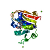
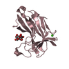
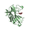
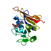
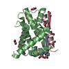
- PDB-2qk8: Crystal structure of the anthrax drug target, Bacillus anthracis ... -
+ Open data

- Basic information
| Entry | Database: PDB / ID: 2qk8 | ||||||
|---|---|---|---|---|---|---|---|
| Title | Crystal structure of the anthrax drug target, Bacillus anthracis dihydrofolate reductase | ||||||
 Components Components | Dihydrofolate reductase | ||||||
 Keywords Keywords | OXIDOREDUCTASE / Methotrexate / Pteridine binding / Nucleotide phosphate binding / Pseudo-Rossman fold | ||||||
| Function / homology |  Function and homology information Function and homology informationglycine biosynthetic process / dihydrofolate reductase / dihydrofolate reductase activity / tetrahydrofolate biosynthetic process / one-carbon metabolic process / NADP binding / metal ion bindingSimilarity search - Function | ||||||
| Biological species |  Bacillus anthracis str. (bacteria) Bacillus anthracis str. (bacteria) | ||||||
| Method | X-RAY DIFFRACTION / SYNCHROTRON / MOLECULAR REPLACEMENT / Resolution: 2.4 Å | ||||||
 Authors Authors | Bennett, B.C. / Xu, H. / Simmerman, R.F. / Lee, R.E. / Dealwis, C.G. | ||||||
 Citation Citation | Journal: J.Med.Chem. / Year: 2007 Title: Crystal structure of the anthrax drug target, Bacillus anthracis dihydrofolate reductase. Authors: Bennett, B.C. / Xu, H. / Simmerman, R.F. / Lee, R.E. / Dealwis, C.G. | ||||||
| History |
|
- Structure visualization
| Structure viewer | Molecule: MolmilJmol/JSmol |
|---|
- Downloads & links
-Download
| PDBx/mmCIF format | 2qk8.cif.gz | 49.1 KB | Display | PDBx/mmCIF format |
|---|---|---|---|---|
| PDB format | pdb2qk8.ent.gz | 34.5 KB | Display | PDB format |
| PDBx/mmJSON format | 2qk8.json.gz | Tree view | PDBx/mmJSON format | |
| Others |  Other downloads Other downloads |
-Validation report
| Arichive directory | https://data.pdbj.org/pub/pdb/validation_reports/qk/2qk8ftp://data.pdbj.org/pub/pdb/validation_reports/qk/2qk8 | HTTPS FTP |
|---|
-Related structure data
| Related structure data | 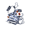 1zdrS S: Starting model for refinement |
|---|---|
| Similar structure data |

-Links
 PDBj
PDBj
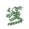
- Assembly
| Deposited unit | 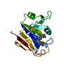
| ||||||||||||
|---|---|---|---|---|---|---|---|---|---|---|---|---|---|
| 1 |
| ||||||||||||
| Unit cell |
| ||||||||||||
| Components on special symmetry positions |
| ||||||||||||
| Details | Only one molecule in the asymmetric unit. Enzyme is functional as a monomer. |
-Components
| #1: Protein | Mass: 19148.854 Da / Num. of mol.: 1 Source method: isolated from a genetically manipulated source Source: (gene. exp.) Bacillus anthracis str. (bacteria) / Species: Bacillus anthracis / Strain: Sterne / Gene: dfrA / Plasmid: Champion pET-SUMO / Species (production host): Escherichia coli / Production host: Escherichia coli BL21(DE3) (bacteria) / Strain (production host): BL21(DE3) / References: UniProt: Q81R22, dihydrofolate reductase |
|---|---|
| #2: Chemical | ChemComp-MTX / Methotrexate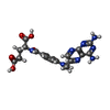  Mass: 454.439 Da / Num. of mol.: 1 / Source method: obtained synthetically / Formula: C20H22N8O5 / Comment: chemotherapy*YM Mass: 454.439 Da / Num. of mol.: 1 / Source method: obtained synthetically / Formula: C20H22N8O5 / Comment: chemotherapy*YM |
| #3: Water | ChemComp-HOH / Water Mass: 18.015 Da / Num. of mol.: 48 / Source method: isolated from a natural source / Formula: H2O Mass: 18.015 Da / Num. of mol.: 48 / Source method: isolated from a natural source / Formula: H2O |
-Experimental details
-Experiment
| Experiment | Method: X-RAY DIFFRACTION / Number of used crystals: 1 |
|---|
- Sample preparation
| Crystal | Density Matthews: 2.6 Å3/Da / Density % sol: 52.76 % |
|---|---|
| Crystal grow | Temperature: 293 K / Method: vapor diffusion, hanging drop / pH: 5.5 Details: 0.1 M Bis-Tris pH 5.5, 0.2 M CaCl2, 20% PEG 3350, VAPOR DIFFUSION, HANGING DROP, temperature 293K |
-Data collection
| Diffraction | Mean temperature: 100 K |
|---|---|
| Diffraction source | Source: SYNCHROTRON / Site: APS  / Beamline: 14-BM-C / Wavelength: 0.9 Å / Beamline: 14-BM-C / Wavelength: 0.9 Å |
| Detector | Type: ADSC QUANTUM 315 / Detector: CCD / Date: Aug 15, 2006 Details: Bent conical Si-mirror (Rh coated) Bent Ge(111) monochromator |
| Radiation | Monochromator: Bent Ge(111) monochromator / Protocol: SINGLE WAVELENGTH / Monochromatic (M) / Laue (L): M / Scattering type: x-ray |
| Radiation wavelength | Wavelength: 0.9 Å / Relative weight: 1 |
| Reflection | Resolution: 2.2→58.4 Å / Num. all: 16860 / Num. obs: 10104 / % possible obs: 92.3 % / Observed criterion σ(I): 2 / Redundancy: 3.3 % / Biso Wilson estimate: 32.8 Å2 / Rsym value: 0.099 / Net I/σ(I): 13.7 |
| Reflection shell | Resolution: 2.2→2.28 Å / Redundancy: 2.9 % / Mean I/σ(I) obs: 2.8 / Rsym value: 0.342 / % possible all: 76.8 |
- Processing
| Software |
| ||||||||||||||||||||||||||||||||||||||||||||||||||||||||||||||||||||||||||||||||||||||||||
|---|---|---|---|---|---|---|---|---|---|---|---|---|---|---|---|---|---|---|---|---|---|---|---|---|---|---|---|---|---|---|---|---|---|---|---|---|---|---|---|---|---|---|---|---|---|---|---|---|---|---|---|---|---|---|---|---|---|---|---|---|---|---|---|---|---|---|---|---|---|---|---|---|---|---|---|---|---|---|---|---|---|---|---|---|---|---|---|---|---|---|---|
| Refinement | Method to determine structure: MOLECULAR REPLACEMENT Starting model: PDB ENTRY: 1ZDR (Bacillus stearothermophilus DHFR) Resolution: 2.4→22 Å / Cor.coef. Fo:Fc: 0.924 / Cor.coef. Fo:Fc free: 0.903 / SU B: 11.98 / SU ML: 0.263 / Isotropic thermal model: Isotropic / Cross valid method: THROUGHOUT / σ(I): 2.8 / ESU R: 0.595 / ESU R Free: 0.303 / Stereochemistry target values: MAXIMUM LIKELIHOOD
| ||||||||||||||||||||||||||||||||||||||||||||||||||||||||||||||||||||||||||||||||||||||||||
| Solvent computation | Ion probe radii: 0.8 Å / Shrinkage radii: 0.8 Å / VDW probe radii: 1.2 Å / Solvent model: MASK | ||||||||||||||||||||||||||||||||||||||||||||||||||||||||||||||||||||||||||||||||||||||||||
| Displacement parameters | Biso mean: 44.402 Å2 | ||||||||||||||||||||||||||||||||||||||||||||||||||||||||||||||||||||||||||||||||||||||||||
| Refinement step | Cycle: LAST / Resolution: 2.4→22 Å
| ||||||||||||||||||||||||||||||||||||||||||||||||||||||||||||||||||||||||||||||||||||||||||
| Refine LS restraints |
| ||||||||||||||||||||||||||||||||||||||||||||||||||||||||||||||||||||||||||||||||||||||||||
| LS refinement shell | Resolution: 2.4→2.462 Å / Total num. of bins used: 20
|
