 Movie
Movie Controller

[English] 日本語
 Yorodumi
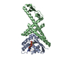
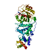
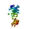
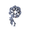
- PDB-2pvs: Structure of human pancreatic lipase related protein 2 mutant N336Q -
+ Open data

- Basic information
| Entry | Database: PDB / ID: 2pvs | ||||||
|---|---|---|---|---|---|---|---|
| Title | Structure of human pancreatic lipase related protein 2 mutant N336Q | ||||||
 Components Components | Pancreatic lipase-related protein 2 | ||||||
 Keywords Keywords |  HYDROLASE / Lipase / galacto lipids hydrolysis HYDROLASE / Lipase / galacto lipids hydrolysis | ||||||
| Function / homology |  Function and homology informationgalactolipase / galactolipid catabolic process / galactolipase activity / 1-18:1-2-16:0-monogalactosyldiacylglycerol lipase activity / Digestion of dietary lipid / lipid digestion / triglyceride catabolic process / phospholipase activity / acylglycerol lipase activity / zymogen granule membrane ...galactolipase / galactolipid catabolic process / galactolipase activity / 1-18:1-2-16:0-monogalactosyldiacylglycerol lipase activity / Digestion of dietary lipid / lipid digestion / triglyceride catabolic process / phospholipase activity / acylglycerol lipase activity / zymogen granule membrane / phospholipid catabolic process / phosphatidylcholine catabolic process / triacylglycerol lipase / triglyceride lipase activity / triglyceride metabolic process / neuron projection / calcium ion binding / extracellular space / extracellular region Function and homology informationgalactolipase / galactolipid catabolic process / galactolipase activity / 1-18:1-2-16:0-monogalactosyldiacylglycerol lipase activity / Digestion of dietary lipid / lipid digestion / triglyceride catabolic process / phospholipase activity / acylglycerol lipase activity / zymogen granule membrane ...galactolipase / galactolipid catabolic process / galactolipase activity / 1-18:1-2-16:0-monogalactosyldiacylglycerol lipase activity / Digestion of dietary lipid / lipid digestion / triglyceride catabolic process / phospholipase activity / acylglycerol lipase activity / zymogen granule membrane / phospholipid catabolic process / phosphatidylcholine catabolic process / triacylglycerol lipase / triglyceride lipase activity / triglyceride metabolic process / neuron projection / calcium ion binding / extracellular space / extracellular regionSimilarity search - Function | ||||||
| Biological species |  Homo sapiens (human) Homo sapiens (human) | ||||||
| Method | X-RAY DIFFRACTION / SYNCHROTRON / MOLECULAR REPLACEMENT / Resolution: 3 Å | ||||||
 Authors Authors | Spinelli, S. / Eydoux, C. / Carriere, F. / Cambillau, C. | ||||||
 Citation Citation | Journal: Biochemistry / Year: 2008 Title: Structure of human pancreatic lipase-related protein 2 with the lid in an open conformation. Authors: Eydoux, C. / Spinelli, S. / Davis, T.L. / Walker, J.R. / Seitova, A. / Dhe-Paganon, S. / De Caro, A. / Cambillau, C. / Carriere, F. | ||||||
| History |
|
- Structure visualization
| Structure viewer | Molecule: MolmilJmol/JSmol |
|---|
- Downloads & links
-Download
| PDBx/mmCIF format | 2pvs.cif.gz | 182.1 KB | Display | PDBx/mmCIF format |
|---|---|---|---|---|
| PDB format | pdb2pvs.ent.gz | 143.9 KB | Display | PDB format |
| PDBx/mmJSON format | 2pvs.json.gz | Tree view | PDBx/mmJSON format | |
| Others |  Other downloads Other downloads |
-Validation report
| Arichive directory | https://data.pdbj.org/pub/pdb/validation_reports/pv/2pvsftp://data.pdbj.org/pub/pdb/validation_reports/pv/2pvs | HTTPS FTP |
|---|
-Related structure data
| Related structure data |  2oxeC  1gplS S: Starting model for refinement C: citing same article ( |
|---|---|
| Similar structure data |


-Links
 PDBj
PDBj- Assembly
| Deposited unit | 
| ||||||||||||||||||||||||||||||||||||||||||||||||||||||||||||||||||||
|---|---|---|---|---|---|---|---|---|---|---|---|---|---|---|---|---|---|---|---|---|---|---|---|---|---|---|---|---|---|---|---|---|---|---|---|---|---|---|---|---|---|---|---|---|---|---|---|---|---|---|---|---|---|---|---|---|---|---|---|---|---|---|---|---|---|---|---|---|---|
| 1 | 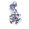
| ||||||||||||||||||||||||||||||||||||||||||||||||||||||||||||||||||||
| 2 | 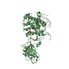
| ||||||||||||||||||||||||||||||||||||||||||||||||||||||||||||||||||||
| 3 | 
| ||||||||||||||||||||||||||||||||||||||||||||||||||||||||||||||||||||
| Unit cell |
| ||||||||||||||||||||||||||||||||||||||||||||||||||||||||||||||||||||
| Noncrystallographic symmetry (NCS) | NCS domain:
NCS domain segments: Component-ID: 1 / Refine code: 5
NCS ensembles :
|
-Components
| #1: Protein | Mass: 50180.184 Da / Num. of mol.: 2 / Mutation: N336Q Source method: isolated from a genetically manipulated source Source: (gene. exp.) Homo sapiens (human) / Gene: PNLIPRP2, PLRP2 / Production host:  Pichia pastoris (fungus) / Strain (production host): SMD 1168 / References: UniProt: P54317, triacylglycerol lipase Pichia pastoris (fungus) / Strain (production host): SMD 1168 / References: UniProt: P54317, triacylglycerol lipase#2: Chemical | ChemComp-SO4 / Sulfate  Mass: 96.063 Da / Num. of mol.: 8 / Source method: obtained synthetically / Formula: SO4 Mass: 96.063 Da / Num. of mol.: 8 / Source method: obtained synthetically / Formula: SO4#3: Chemical |   Mass: 40.078 Da / Num. of mol.: 2 / Source method: obtained synthetically / Formula: Ca Mass: 40.078 Da / Num. of mol.: 2 / Source method: obtained synthetically / Formula: Ca#4: Water | ChemComp-HOH / | Water Mass: 18.015 Da / Num. of mol.: 67 / Source method: isolated from a natural source / Formula: H2O Mass: 18.015 Da / Num. of mol.: 67 / Source method: isolated from a natural source / Formula: H2O |
|---|
-Experimental details
-Experiment
| Experiment | Method: X-RAY DIFFRACTION / Number of used crystals: 1 |
|---|
- Sample preparation
| Crystal | Density Matthews: 3.6 Å3/Da / Density % sol: 65.81 % |
|---|---|
| Crystal grow | Temperature: 293 K / Method: vapor diffusion, hanging drop / pH: 6.7 Details: 6 microliter of protein (14.5 mg/ml in 0.2 M NaCl, 25 mM Tris-HCl, pH 8.0) and 2 microliter of 2.05 M Ammonium sulphate, 0.1 M Hepes, pH 6.7, VAPOR DIFFUSION, HANGING DROP, temperature 293K |
-Data collection
| Diffraction | Mean temperature: 100 K |
|---|---|
| Diffraction source | Source: SYNCHROTRON / Site: ESRF  / Beamline: ID14-3 / Wavelength: 0.931 / Beamline: ID14-3 / Wavelength: 0.931 |
| Detector | Type: ADSC QUANTUM 4 / Detector: CCD / Date: Feb 9, 2007 / Details: mirrors |
| Radiation | Protocol: SINGLE WAVELENGTH / Monochromatic (M) / Laue (L): M / Scattering type: x-ray |
| Radiation wavelength | Wavelength: 0.931 Å / Relative weight: 1 |
| Reflection | Resolution: 3→35 Å / Num. all: 27791 / Num. obs: 27791 / % possible obs: 100 % / Observed criterion σ(F): 0 / Observed criterion σ(I): 0 / Redundancy: 6 % / Biso Wilson estimate: 88 Å2 / Rmerge(I) obs: 0.118 / Rsym value: 0.118 / Net I/σ(I): 10.5 |
| Reflection shell | Resolution: 3→3.16 Å / Redundancy: 6.1 % / Rmerge(I) obs: 0.48 / Mean I/σ(I) obs: 1.5 / Num. unique all: 4339 / Rsym value: 0.48 / % possible all: 100 |
- Processing
| Software |
| ||||||||||||||||||||||||||||||||||||||||||||||||||||||||||||||||||||||||||||||||||||||||||||||||||||||||||||||||||||||||||||||||||
|---|---|---|---|---|---|---|---|---|---|---|---|---|---|---|---|---|---|---|---|---|---|---|---|---|---|---|---|---|---|---|---|---|---|---|---|---|---|---|---|---|---|---|---|---|---|---|---|---|---|---|---|---|---|---|---|---|---|---|---|---|---|---|---|---|---|---|---|---|---|---|---|---|---|---|---|---|---|---|---|---|---|---|---|---|---|---|---|---|---|---|---|---|---|---|---|---|---|---|---|---|---|---|---|---|---|---|---|---|---|---|---|---|---|---|---|---|---|---|---|---|---|---|---|---|---|---|---|---|---|---|---|
| Refinement | Method to determine structure: MOLECULAR REPLACEMENT Starting model: pdb entry 1GPL Resolution: 3→34.18 Å / Cor.coef. Fo:Fc: 0.936 / Cor.coef. Fo:Fc free: 0.888 / SU B: 31.566 / SU ML: 0.279 / Cross valid method: THROUGHOUT / σ(F): 0 / σ(I): 0 / ESU R Free: 0.383 / Stereochemistry target values: MAXIMUM LIKELIHOOD / Details: HYDROGENS HAVE BEEN ADDED IN THE RIDING POSITIONS
| ||||||||||||||||||||||||||||||||||||||||||||||||||||||||||||||||||||||||||||||||||||||||||||||||||||||||||||||||||||||||||||||||||
| Solvent computation | Ion probe radii: 0.8 Å / Shrinkage radii: 0.8 Å / VDW probe radii: 1.2 Å / Solvent model: MASK | ||||||||||||||||||||||||||||||||||||||||||||||||||||||||||||||||||||||||||||||||||||||||||||||||||||||||||||||||||||||||||||||||||
| Displacement parameters | Biso mean: 51.447 Å2
| ||||||||||||||||||||||||||||||||||||||||||||||||||||||||||||||||||||||||||||||||||||||||||||||||||||||||||||||||||||||||||||||||||
| Refine analyze | Luzzati coordinate error free: 0.38 Å | ||||||||||||||||||||||||||||||||||||||||||||||||||||||||||||||||||||||||||||||||||||||||||||||||||||||||||||||||||||||||||||||||||
| Refinement step | Cycle: LAST / Resolution: 3→34.18 Å
| ||||||||||||||||||||||||||||||||||||||||||||||||||||||||||||||||||||||||||||||||||||||||||||||||||||||||||||||||||||||||||||||||||
| Refine LS restraints |
| ||||||||||||||||||||||||||||||||||||||||||||||||||||||||||||||||||||||||||||||||||||||||||||||||||||||||||||||||||||||||||||||||||
| Refine LS restraints NCS | Dom-ID: 1 / Auth asym-ID: A / Refine-ID: X-RAY DIFFRACTION
| ||||||||||||||||||||||||||||||||||||||||||||||||||||||||||||||||||||||||||||||||||||||||||||||||||||||||||||||||||||||||||||||||||
| LS refinement shell | Resolution: 3→3.077 Å / Total num. of bins used: 20
|
