 Movie
Movie Controller


+ Open data

- Basic information
| Entry | Database: PDB / ID: 3feo | ||||||
|---|---|---|---|---|---|---|---|
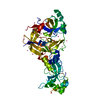
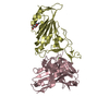
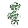
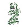
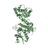
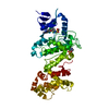
| Title | The crystal structure of MBTD1 | ||||||
 Components Components | MBT domain-containing protein 1 | ||||||
 Keywords Keywords | METAL BINDING PROTEIN / MBTL1 /  Structural Genomics / Structural Genomics Consortium / SGC / Metal-binding / Nucleus / Zinc-finger Structural Genomics / Structural Genomics Consortium / SGC / Metal-binding / Nucleus / Zinc-finger | ||||||
| Function / homology |  Function and homology informationNuA4 histone acetyltransferase complex binding / embryonic skeletal system development / regulation of double-strand break repair / NuA4 histone acetyltransferase complex / positive regulation of double-strand break repair via homologous recombination / methylated histone binding / double-strand break repair via homologous recombination / nucleosome / site of double-strand break / chromatin organization ...NuA4 histone acetyltransferase complex binding / embryonic skeletal system development / regulation of double-strand break repair / NuA4 histone acetyltransferase complex / positive regulation of double-strand break repair via homologous recombination / methylated histone binding / double-strand break repair via homologous recombination / nucleosome / site of double-strand break / chromatin organization / regulation of apoptotic process / regulation of cell cycle / negative regulation of DNA-templated transcription / chromatin binding / positive regulation of DNA-templated transcription / zinc ion binding / nucleus Function and homology informationNuA4 histone acetyltransferase complex binding / embryonic skeletal system development / regulation of double-strand break repair / NuA4 histone acetyltransferase complex / positive regulation of double-strand break repair via homologous recombination / methylated histone binding / double-strand break repair via homologous recombination / nucleosome / site of double-strand break / chromatin organization ...NuA4 histone acetyltransferase complex binding / embryonic skeletal system development / regulation of double-strand break repair / NuA4 histone acetyltransferase complex / positive regulation of double-strand break repair via homologous recombination / methylated histone binding / double-strand break repair via homologous recombination / nucleosome / site of double-strand break / chromatin organization / regulation of apoptotic process / regulation of cell cycle / negative regulation of DNA-templated transcription / chromatin binding / positive regulation of DNA-templated transcription / zinc ion binding / nucleusSimilarity search - Function | ||||||
| Biological species |  Homo sapiens (human) Homo sapiens (human) | ||||||
| Method | X-RAY DIFFRACTION / SYNCHROTRON / MOLECULAR REPLACEMENT / Resolution: 2.5 Å | ||||||
 Authors Authors | Amaya, M.F. / Eryilmaz, J. / Kozieradzki, I. / Edwards, A.M. / Arrowsmith, C.H. / Weigelt, J. / Bountra, C. / Bochkarev, A. / Min, J. / Structural Genomics Consortium (SGC) | ||||||
 Citation Citation | Journal: Plos One / Year: 2009 Title: Structural studies of a four-MBT repeat protein MBTD1. Authors: Eryilmaz, J. / Pan, P. / Amaya, M.F. / Allali-Hassani, A. / Dong, A. / Adams-Cioaba, M.A. / Mackenzie, F. / Vedadi, M. / Min, J. | ||||||
| History |
|
- Structure visualization
| Structure viewer | Molecule: MolmilJmol/JSmol |
|---|
- Downloads & links
-Download
| PDBx/mmCIF format | 3feo.cif.gz | 166.8 KB | Display | PDBx/mmCIF format |
|---|---|---|---|---|
| PDB format | pdb3feo.ent.gz | 130.6 KB | Display | PDB format |
| PDBx/mmJSON format | 3feo.json.gz | Tree view | PDBx/mmJSON format | |
| Others |  Other downloads Other downloads |
-Validation report
| Arichive directory | https://data.pdbj.org/pub/pdb/validation_reports/fe/3feoftp://data.pdbj.org/pub/pdb/validation_reports/fe/3feo | HTTPS FTP |
|---|
-Related structure data
| Related structure data | 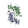 3f70S S: Starting model for refinement |
|---|---|
| Similar structure data |


-Links
 PDBj
PDBj- Assembly
| Deposited unit | 
| ||||||||
|---|---|---|---|---|---|---|---|---|---|
| 1 | 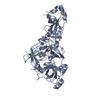
| ||||||||
| 2 | 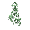
| ||||||||
| Unit cell |
|
-Components
| #1: Protein | Mass: 49565.723 Da / Num. of mol.: 2 Source method: isolated from a genetically manipulated source Source: (gene. exp.) Homo sapiens (human) / Gene: MBTD1 / Production host:  Escherichia coli (E. coli) / References: UniProt: Q05BQ5 Escherichia coli (E. coli) / References: UniProt: Q05BQ5#2: Water | ChemComp-HOH / | Water Mass: 18.015 Da / Num. of mol.: 71 / Source method: isolated from a natural source / Formula: H2O Mass: 18.015 Da / Num. of mol.: 71 / Source method: isolated from a natural source / Formula: H2O |
|---|
-Experimental details
-Experiment
| Experiment | Method: X-RAY DIFFRACTION / Number of used crystals: 1 |
|---|
- Sample preparation
| Crystal | Density Matthews: 2.42 Å3/Da / Density % sol: 49.19 % |
|---|---|
| Crystal grow | Method: vapor diffusion, hanging drop / pH: 7.5 Details: 0.2M CaOAc, 20% PEG3350, pH 7.5, VAPOR DIFFUSION, HANGING DROP |
-Data collection
| Diffraction | Mean temperature: 200 K | |||||||||||||||||||||||||||||||||||||||||||||||||||||||||||||||||||||||||||||
|---|---|---|---|---|---|---|---|---|---|---|---|---|---|---|---|---|---|---|---|---|---|---|---|---|---|---|---|---|---|---|---|---|---|---|---|---|---|---|---|---|---|---|---|---|---|---|---|---|---|---|---|---|---|---|---|---|---|---|---|---|---|---|---|---|---|---|---|---|---|---|---|---|---|---|---|---|---|---|
| Diffraction source | Source: SYNCHROTRON / Site: APS  / Beamline: 23-ID-B / Wavelength: 0.97948 Å / Beamline: 23-ID-B / Wavelength: 0.97948 Å | |||||||||||||||||||||||||||||||||||||||||||||||||||||||||||||||||||||||||||||
| Detector | Date: Nov 22, 2008 | |||||||||||||||||||||||||||||||||||||||||||||||||||||||||||||||||||||||||||||
| Radiation | Protocol: SINGLE WAVELENGTH / Monochromatic (M) / Laue (L): M / Scattering type: x-ray | |||||||||||||||||||||||||||||||||||||||||||||||||||||||||||||||||||||||||||||
| Radiation wavelength | Wavelength: 0.97948 Å / Relative weight: 1 | |||||||||||||||||||||||||||||||||||||||||||||||||||||||||||||||||||||||||||||
| Reflection | Resolution: 2.5→100 Å / Num. obs: 34002 / % possible obs: 99 % / Redundancy: 6.9 % / Rmerge(I) obs: 0.091 / Χ2: 1.529 / Net I/σ(I): 26.317 | |||||||||||||||||||||||||||||||||||||||||||||||||||||||||||||||||||||||||||||
| Reflection shell |
|
- Processing
| Software |
| |||||||||||||||||||||||||||||||||||||||||||||||||||||||||||||||||||||||||||||||||||||||||||||||||||||||||||||||||||||||||||||||||||||||||||||||||||||||||||||||||||||||||||||||
|---|---|---|---|---|---|---|---|---|---|---|---|---|---|---|---|---|---|---|---|---|---|---|---|---|---|---|---|---|---|---|---|---|---|---|---|---|---|---|---|---|---|---|---|---|---|---|---|---|---|---|---|---|---|---|---|---|---|---|---|---|---|---|---|---|---|---|---|---|---|---|---|---|---|---|---|---|---|---|---|---|---|---|---|---|---|---|---|---|---|---|---|---|---|---|---|---|---|---|---|---|---|---|---|---|---|---|---|---|---|---|---|---|---|---|---|---|---|---|---|---|---|---|---|---|---|---|---|---|---|---|---|---|---|---|---|---|---|---|---|---|---|---|---|---|---|---|---|---|---|---|---|---|---|---|---|---|---|---|---|---|---|---|---|---|---|---|---|---|---|---|---|---|---|---|---|---|
| Refinement | Method to determine structure: MOLECULAR REPLACEMENT Starting model: PDB entry 3F70 Resolution: 2.5→80.85 Å / Cor.coef. Fo:Fc: 0.932 / Cor.coef. Fo:Fc free: 0.888 / Occupancy max: 1 / Occupancy min: 0.5 / SU B: 20.504 / SU ML: 0.218 / TLS residual ADP flag: LIKELY RESIDUAL / Cross valid method: THROUGHOUT / σ(F): 0 / ESU R: 0.497 / ESU R Free: 0.308 / Stereochemistry target values: MAXIMUM LIKELIHOOD / Details: HYDROGENS HAVE BEEN ADDED IN THE RIDING POSITIONS
| |||||||||||||||||||||||||||||||||||||||||||||||||||||||||||||||||||||||||||||||||||||||||||||||||||||||||||||||||||||||||||||||||||||||||||||||||||||||||||||||||||||||||||||||
| Solvent computation | Ion probe radii: 0.8 Å / Shrinkage radii: 0.8 Å / VDW probe radii: 1.2 Å / Solvent model: MASK | |||||||||||||||||||||||||||||||||||||||||||||||||||||||||||||||||||||||||||||||||||||||||||||||||||||||||||||||||||||||||||||||||||||||||||||||||||||||||||||||||||||||||||||||
| Displacement parameters | Biso max: 55.15 Å2 / Biso mean: 21.071 Å2 / Biso min: 2 Å2
| |||||||||||||||||||||||||||||||||||||||||||||||||||||||||||||||||||||||||||||||||||||||||||||||||||||||||||||||||||||||||||||||||||||||||||||||||||||||||||||||||||||||||||||||
| Refinement step | Cycle: LAST / Resolution: 2.5→80.85 Å
| |||||||||||||||||||||||||||||||||||||||||||||||||||||||||||||||||||||||||||||||||||||||||||||||||||||||||||||||||||||||||||||||||||||||||||||||||||||||||||||||||||||||||||||||
| Refine LS restraints |
| |||||||||||||||||||||||||||||||||||||||||||||||||||||||||||||||||||||||||||||||||||||||||||||||||||||||||||||||||||||||||||||||||||||||||||||||||||||||||||||||||||||||||||||||
| LS refinement shell | Resolution: 2.5→2.558 Å / Total num. of bins used: 20
| |||||||||||||||||||||||||||||||||||||||||||||||||||||||||||||||||||||||||||||||||||||||||||||||||||||||||||||||||||||||||||||||||||||||||||||||||||||||||||||||||||||||||||||||
| Refinement TLS params. | Method: refined / Refine-ID: X-RAY DIFFRACTION
| |||||||||||||||||||||||||||||||||||||||||||||||||||||||||||||||||||||||||||||||||||||||||||||||||||||||||||||||||||||||||||||||||||||||||||||||||||||||||||||||||||||||||||||||
| Refinement TLS group |
|
