 Movie
Movie Controller


+ Open data

- Basic information
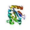
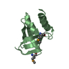
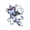
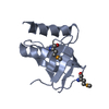
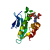
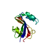
| Entry | Database: PDB / ID: 2pko | ||||||
|---|---|---|---|---|---|---|---|
| Title | Crystal structure of yeast Urm1 at 1.8 A resolution | ||||||
 Components Components | Ubiquitin-related modifier 1 | ||||||
 Keywords Keywords |  PROTEIN BINDING / Beta Grasp Fold Ubiquitin-like protein PROTEIN BINDING / Beta Grasp Fold Ubiquitin-like protein | ||||||
| Function / homology |  Function and homology information Function and homology informationcell budding / tRNA thio-modification / sulfur carrier activity / tRNA wobble position uridine thiolation / protein urmylation / invasive growth in response to glucose limitation / tRNA wobble uridine modification / protein tag activity / cellular response to oxidative stress / protein homodimerization activity ...cell budding / tRNA thio-modification / sulfur carrier activity / tRNA wobble position uridine thiolation / protein urmylation / invasive growth in response to glucose limitation / tRNA wobble uridine modification / protein tag activity / cellular response to oxidative stress / protein homodimerization activity / nucleus / cytosol / cytoplasmSimilarity search - Function | ||||||
| Biological species |  Saccharomyces cerevisiae (brewer's yeast) Saccharomyces cerevisiae (brewer's yeast) | ||||||
| Method | X-RAY DIFFRACTION / SYNCHROTRON / MOLECULAR REPLACEMENT / Resolution: 1.8 Å | ||||||
 Authors Authors | Lee, E.Y. / Lake, M.W. / Schindelin, H. | ||||||
 Citation Citation | Journal: Biochemistry / Year: 2008 Title: The sulfurtransferase activity of Uba4 presents a link between ubiquitin-like protein conjugation and activation of sulfur carrier proteins. Authors: Schmitz, J. / Chowdhury, M.M. / Hanzelmann, P. / Nimtz, M. / Lee, E.Y. / Schindelin, H. / Leimkuhler, S. | ||||||
| History |
|
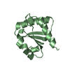
- Structure visualization
| Structure viewer | Molecule: MolmilJmol/JSmol |
|---|
- Downloads & links
-Download
| PDBx/mmCIF format | 2pko.cif.gz | 33.9 KB | Display | PDBx/mmCIF format |
|---|---|---|---|---|
| PDB format | pdb2pko.ent.gz | 22.7 KB | Display | PDB format |
| PDBx/mmJSON format | 2pko.json.gz | Tree view | PDBx/mmJSON format | |
| Others |  Other downloads Other downloads |
-Validation report
| Arichive directory | https://data.pdbj.org/pub/pdb/validation_reports/pk/2pkoftp://data.pdbj.org/pub/pdb/validation_reports/pk/2pko | HTTPS FTP |
|---|
-Related structure data
| Related structure data |  1xo3S S: Starting model for refinement |
|---|---|
| Similar structure data |

-Links
 PDBj
PDBj


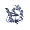
- Assembly
| Deposited unit | 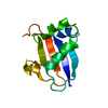
| ||||||||
|---|---|---|---|---|---|---|---|---|---|
| 1 |
| ||||||||
| Unit cell |
| ||||||||
| Components on special symmetry positions |
| ||||||||
| Details | Monomer with one molecule in the asymetric unit |
-Components
| #1: Protein | Mass: 11039.428 Da / Num. of mol.: 1 Source method: isolated from a genetically manipulated source Source: (gene. exp.) Saccharomyces cerevisiae (brewer's yeast)Gene: URM1 / Plasmid: pET28a / Species (production host): Escherichia coli / Production host:  Escherichia coli BL21(DE3) (bacteria) / Strain (production host): BL21 DE3 / References: UniProt: P40554 Escherichia coli BL21(DE3) (bacteria) / Strain (production host): BL21 DE3 / References: UniProt: P40554 |
|---|---|
| #2: Water | ChemComp-HOH / Water Mass: 18.015 Da / Num. of mol.: 79 / Source method: isolated from a natural source / Formula: H2O Mass: 18.015 Da / Num. of mol.: 79 / Source method: isolated from a natural source / Formula: H2O |
-Experimental details
-Experiment
| Experiment | Method: X-RAY DIFFRACTION / Number of used crystals: 1 |
|---|
- Sample preparation
| Crystal | Density Matthews: 1.97 Å3/Da / Density % sol: 37.46 % |
|---|---|
| Crystal grow | Temperature: 291 K / Method: vapor diffusion, hanging drop / pH: 6.5 Details: 33-35% PEG 8000, 0.1M sodium cacodylate, pH 6.5, VAPOR DIFFUSION, HANGING DROP, temperature 291K |
-Data collection
| Diffraction | Mean temperature: 100 K |
|---|---|
| Diffraction source | Source: SYNCHROTRON / Site: NSLS  / Beamline: X26C / Wavelength: 1.1 Å / Beamline: X26C / Wavelength: 1.1 Å |
| Detector | Type: ADSC QUANTUM 4 / Detector: CCD / Date: May 29, 2003 |
| Radiation | Monochromator: channel-cut Si(111) / Protocol: SINGLE WAVELENGTH / Monochromatic (M) / Laue (L): M / Scattering type: x-ray |
| Radiation wavelength | Wavelength: 1.1 Å / Relative weight: 1 |
| Reflection | Resolution: 1.8→19.75 Å / Num. all: 7848 / Num. obs: 7848 / % possible obs: 97.16 % / Observed criterion σ(F): 0 / Observed criterion σ(I): -3 |
| Reflection shell | Highest resolution: 1.8 Å / % possible all: 97.16 |
- Processing
| Software |
| ||||||||||||||||||||||||||||||||||||||||||||||||||||||||||||||||||||||||||||||||||||||||||||||||||||||||||||||||||||||||||||||||||
|---|---|---|---|---|---|---|---|---|---|---|---|---|---|---|---|---|---|---|---|---|---|---|---|---|---|---|---|---|---|---|---|---|---|---|---|---|---|---|---|---|---|---|---|---|---|---|---|---|---|---|---|---|---|---|---|---|---|---|---|---|---|---|---|---|---|---|---|---|---|---|---|---|---|---|---|---|---|---|---|---|---|---|---|---|---|---|---|---|---|---|---|---|---|---|---|---|---|---|---|---|---|---|---|---|---|---|---|---|---|---|---|---|---|---|---|---|---|---|---|---|---|---|---|---|---|---|---|---|---|---|---|
| Refinement | Method to determine structure: MOLECULAR REPLACEMENT Starting model: PDB ENTRY 1XO3 Resolution: 1.8→19.75 Å / Cor.coef. Fo:Fc: 0.958 / Cor.coef. Fo:Fc free: 0.929 / SU B: 3.727 / SU ML: 0.116 / Cross valid method: THROUGHOUT / σ(F): 0 / ESU R: 0.157 / ESU R Free: 0.163 / Stereochemistry target values: MAXIMUM LIKELIHOOD / Details: HYDROGENS HAVE BEEN ADDED IN THE RIDING POSITIONS
| ||||||||||||||||||||||||||||||||||||||||||||||||||||||||||||||||||||||||||||||||||||||||||||||||||||||||||||||||||||||||||||||||||
| Solvent computation | Ion probe radii: 0.8 Å / Shrinkage radii: 0.8 Å / VDW probe radii: 1.2 Å / Solvent model: BABINET MODEL WITH MASK | ||||||||||||||||||||||||||||||||||||||||||||||||||||||||||||||||||||||||||||||||||||||||||||||||||||||||||||||||||||||||||||||||||
| Displacement parameters | Biso mean: 25.252 Å2
| ||||||||||||||||||||||||||||||||||||||||||||||||||||||||||||||||||||||||||||||||||||||||||||||||||||||||||||||||||||||||||||||||||
| Refine analyze |
| ||||||||||||||||||||||||||||||||||||||||||||||||||||||||||||||||||||||||||||||||||||||||||||||||||||||||||||||||||||||||||||||||||
| Refinement step | Cycle: LAST / Resolution: 1.8→19.75 Å
| ||||||||||||||||||||||||||||||||||||||||||||||||||||||||||||||||||||||||||||||||||||||||||||||||||||||||||||||||||||||||||||||||||
| Refine LS restraints |
| ||||||||||||||||||||||||||||||||||||||||||||||||||||||||||||||||||||||||||||||||||||||||||||||||||||||||||||||||||||||||||||||||||
| LS refinement shell | Resolution: 1.804→1.851 Å / Total num. of bins used: 20
|
