 Movie
Movie Controller

[English] 日本語
 Yorodumi
- PDB-3dsg: XC1028 from Xanthomonas campestris Adopts a PilZ Domain-like Stru... -
+ Open data

- Basic information
| Entry | Database: PDB / ID: 3dsg | ||||||
|---|---|---|---|---|---|---|---|
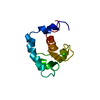
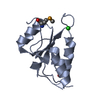
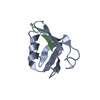
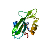
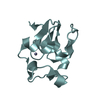
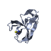
| Title | XC1028 from Xanthomonas campestris Adopts a PilZ Domain-like Structure Yet with Trivial c-di-GMP Binding Activity | ||||||
 Components Components | Type IV fimbriae assembly protein | ||||||
 Keywords Keywords | UNKNOWN FUNCTION /  PilZ domain / Xanthomonas campestris / c-di-GMP / type IV pilus / PA2960 PilZ domain / Xanthomonas campestris / c-di-GMP / type IV pilus / PA2960 | ||||||
| Function / homology | predicted glycosyltransferase like domains / PilZ domain / PilZ domain / cyclic-di-GMP binding / Thrombin, subunit H / Beta Barrel / Mainly Beta / Type IV fimbriae assembly protein Function and homology information Function and homology information | ||||||
| Biological species |  Xanthomonas campestris pv. campestris (bacteria) Xanthomonas campestris pv. campestris (bacteria) | ||||||
| Method | X-RAY DIFFRACTION / SYNCHROTRON / MAD / Resolution: 2.09 Å | ||||||
 Authors Authors | Li, T.N. / Chin, K.H. / Liu, J.H. / Wang, A.H.J. / Chou, S.H. | ||||||
 Citation Citation | Journal: Proteins / Year: 2009 Title: XC1028 from Xanthomonas campestris adopts a PilZ domain-like structure without a c-di-GMP switch. Authors: Li, T.N. / Chin, K.H. / Liu, J.H. / Wang, A.H. / Chou, S.H. | ||||||
| History |
|
- Structure visualization
| Structure viewer | Molecule: MolmilJmol/JSmol |
|---|
- Downloads & links
-Download
| PDBx/mmCIF format | 3dsg.cif.gz | 65.2 KB | Display | PDBx/mmCIF format |
|---|---|---|---|---|
| PDB format | pdb3dsg.ent.gz | 53.2 KB | Display | PDB format |
| PDBx/mmJSON format | 3dsg.json.gz | Tree view | PDBx/mmJSON format | |
| Others |  Other downloads Other downloads |
-Validation report
| Arichive directory | https://data.pdbj.org/pub/pdb/validation_reports/ds/3dsgftp://data.pdbj.org/pub/pdb/validation_reports/ds/3dsg | HTTPS FTP |
|---|
-Related structure data
| Similar structure data |
|---|


-Links
 PDBj
PDBj

- Assembly
| Deposited unit | 
| ||||||||
|---|---|---|---|---|---|---|---|---|---|
| 1 | 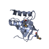
| ||||||||
| 2 | 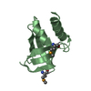
| ||||||||
| 3 | 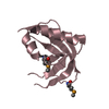
| ||||||||
| Unit cell |
| ||||||||
| Details | C2 |
-Components
| #1: Protein | Mass: 10548.862 Da / Num. of mol.: 3 / Fragment: PilZ domain Source method: isolated from a genetically manipulated source Source: (gene. exp.) Xanthomonas campestris pv. campestris (bacteria)Production host: Escherichia coli (E. coli) / References: UniProt: Q8PBU4#2: Water | ChemComp-HOH / | Water Mass: 18.015 Da / Num. of mol.: 126 / Source method: isolated from a natural source / Formula: H2O Mass: 18.015 Da / Num. of mol.: 126 / Source method: isolated from a natural source / Formula: H2O |
|---|
-Experimental details
-Experiment
| Experiment | Method: X-RAY DIFFRACTION / Number of used crystals: 1 |
|---|
- Sample preparation
| Crystal | Density Matthews: 3.28 Å3/Da / Density % sol: 62.52 % |
|---|---|
| Crystal grow | Temperature: 297 K / Method: vapor diffusion / pH: 6.5 Details: 0.1M sodium cacodylate pH 6.5, 18% PEG 2KMME, 2% glycerol, VAPOR DIFFUSION, temperature 297K |
-Data collection
| Diffraction | Mean temperature: 100 K | ||||||||||||
|---|---|---|---|---|---|---|---|---|---|---|---|---|---|
| Diffraction source | Source: SYNCHROTRON / Site: NSRRC  / Beamline: BL13B1 / Wavelength: 0.97920, 0.96398, 0.97888 / Beamline: BL13B1 / Wavelength: 0.97920, 0.96398, 0.97888 | ||||||||||||
| Detector | Type: ADSC QUANTUM 315 / Detector: CCD / Date: Jan 5, 2008 | ||||||||||||
| Radiation | Protocol: MAD / Monochromatic (M) / Laue (L): M / Scattering type: x-ray | ||||||||||||
| Radiation wavelength |
| ||||||||||||
| Reflection | Resolution: 2.09→30 Å / Num. obs: 22930 / % possible obs: 94.7 % / Observed criterion σ(F): 2 / Observed criterion σ(I): 2 / Redundancy: 5.7 % / Biso Wilson estimate: 21.5 Å2 / Rmerge(I) obs: 0.044 / Rsym value: 0.22 / Net I/σ(I): 27.3 | ||||||||||||
| Reflection shell | Resolution: 2.1→2.18 Å / Redundancy: 4.4 % / Rmerge(I) obs: 0.22 / Mean I/σ(I) obs: 5.3 / Num. unique all: 1714 / Rsym value: 0.23 / % possible all: 79 |
- Processing
| Software |
| ||||||||||||||||||||||||||||||||||||||||||||||||||||||||||||||||||||||||||||||||
|---|---|---|---|---|---|---|---|---|---|---|---|---|---|---|---|---|---|---|---|---|---|---|---|---|---|---|---|---|---|---|---|---|---|---|---|---|---|---|---|---|---|---|---|---|---|---|---|---|---|---|---|---|---|---|---|---|---|---|---|---|---|---|---|---|---|---|---|---|---|---|---|---|---|---|---|---|---|---|---|---|---|
| Refinement | Method to determine structure: MAD / Resolution: 2.09→25.48 Å / Rfactor Rfree error: 0.005 / Data cutoff high absF: 235091.45 / Data cutoff low absF: 0 / Isotropic thermal model: RESTRAINED / Cross valid method: THROUGHOUT / σ(F): 0 / Details: BULK SOLVENT MODEL USED
| ||||||||||||||||||||||||||||||||||||||||||||||||||||||||||||||||||||||||||||||||
| Solvent computation | Solvent model: FLAT MODEL / Bsol: 68.691 Å2 / ksol: 0.4 e/Å3 | ||||||||||||||||||||||||||||||||||||||||||||||||||||||||||||||||||||||||||||||||
| Displacement parameters | Biso mean: 58.3 Å2
| ||||||||||||||||||||||||||||||||||||||||||||||||||||||||||||||||||||||||||||||||
| Refine analyze |
| ||||||||||||||||||||||||||||||||||||||||||||||||||||||||||||||||||||||||||||||||
| Refinement step | Cycle: LAST / Resolution: 2.09→25.48 Å
| ||||||||||||||||||||||||||||||||||||||||||||||||||||||||||||||||||||||||||||||||
| Refine LS restraints |
| ||||||||||||||||||||||||||||||||||||||||||||||||||||||||||||||||||||||||||||||||
| LS refinement shell | Resolution: 2→2.13 Å / Total num. of bins used: 6
| ||||||||||||||||||||||||||||||||||||||||||||||||||||||||||||||||||||||||||||||||
| Xplor file |
|
