 Movie
Movie Controller

[English] 日本語
 Yorodumi
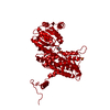
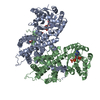
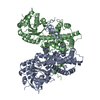
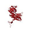
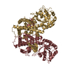
- PDB-2pgd: THE STRUCTURE OF 6-PHOSPHOGLUCONATE DEHYDROGENASE REFINED AT 2 AN... -
+ Open data

- Basic information
| Entry | Database: PDB / ID: 2pgd | |||||||||
|---|---|---|---|---|---|---|---|---|---|---|
| Title | THE STRUCTURE OF 6-PHOSPHOGLUCONATE DEHYDROGENASE REFINED AT 2 ANGSTROMS RESOLUTION | |||||||||
 Components Components | 6-PHOSPHOGLUCONATE DEHYDROGENASE | |||||||||
 Keywords Keywords | OXIDOREDUCTASE (CHOH(D)-NADP+(A)) | |||||||||
| Function / homology |  Function and homology information Function and homology informationD-gluconate metabolic process / phosphogluconate dehydrogenase (NADP+-dependent, decarboxylating) / phosphogluconate dehydrogenase (decarboxylating) activity / pentose-phosphate shunt / NADP binding / cytoplasmSimilarity search - Function | |||||||||
| Biological species |  Ovis aries (sheep) Ovis aries (sheep) | |||||||||
| Method | X-RAY DIFFRACTION / Resolution: 2 Å | |||||||||
 Authors Authors | Adams, M.J. / Phillips, C. / Gover, S. | |||||||||
 Citation Citation | Journal: Acta Crystallogr.,Sect.B / Year: 1991 Title: The structure of 6-phosphogluconate dehydrogenase refined at 2.5 A resolution. Authors: Adams, M.J. / Gover, S. / Leaback, R. / Phillips, C. / Somers, D.O. #1: Journal: Structure / Year: 1994Title: Crystallographic Study of Coenzyme, Coenzyme Analogue and Substrate Binding in 6-Phosphogluconate Dehydrogenase: Implications for Nadp Specificity and the Enzyme Mechanism Authors: Adams, M.J. / Ellis, G.H. / Gover, S. / Naylor, C.E. / Phillips, C. #2: Journal: Acta Crystallogr.,Sect.B / Year: 1991Title: The Structure of 6-Phosphogluconate Dehydrogenase Refined at 2.5 Angstroms Resolution Authors: Adams, M.J. / Gover, S. / Leaback, R. / Phillips, C. / Somers, D.O'N. | |||||||||
| History |
|
- Structure visualization
| Structure viewer | Molecule: MolmilJmol/JSmol |
|---|
- Downloads & links
-Download
| PDBx/mmCIF format | 2pgd.cif.gz | 109.2 KB | Display | PDBx/mmCIF format |
|---|---|---|---|---|
| PDB format | pdb2pgd.ent.gz | 84.5 KB | Display | PDB format |
| PDBx/mmJSON format | 2pgd.json.gz | Tree view | PDBx/mmJSON format | |
| Others |  Other downloads Other downloads |
-Validation report
| Arichive directory | https://data.pdbj.org/pub/pdb/validation_reports/pg/2pgdftp://data.pdbj.org/pub/pdb/validation_reports/pg/2pgd | HTTPS FTP |
|---|
-Related structure data
| Similar structure data |
|---|


-Links
 PDBj
PDBj


- Assembly
| Deposited unit | 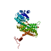
| ||||||||
|---|---|---|---|---|---|---|---|---|---|
| 1 | 
| ||||||||
| Unit cell |
| ||||||||
| Atom site foot note | 1: CIS PROLINE - PRO 67 |
-Components
| #1: Protein | Mass: 52905.652 Da / Num. of mol.: 1 Source method: isolated from a genetically manipulated source Source: (gene. exp.) Ovis aries (sheep)References: UniProt: P00349, phosphogluconate dehydrogenase (NADP+-dependent, decarboxylating) | ||
|---|---|---|---|
| #2: Chemical | Sulfate  Mass: 96.063 Da / Num. of mol.: 3 / Source method: obtained synthetically / Formula: SO4 Mass: 96.063 Da / Num. of mol.: 3 / Source method: obtained synthetically / Formula: SO4#3: Water | ChemComp-HOH / | Water Mass: 18.015 Da / Num. of mol.: 346 / Source method: isolated from a natural source / Formula: H2O Mass: 18.015 Da / Num. of mol.: 346 / Source method: isolated from a natural source / Formula: H2O |
-Experimental details
-Experiment
| Experiment | Method: X-RAY DIFFRACTION |
|---|
- Sample preparation
| Crystal | Density Matthews: 2.61 Å3/Da / Density % sol: 52.87 % | |||||||||||||||||||||||||
|---|---|---|---|---|---|---|---|---|---|---|---|---|---|---|---|---|---|---|---|---|---|---|---|---|---|---|
| Crystal grow | *PLUS pH: 6.5 / Method: vapor diffusion, hanging drop | |||||||||||||||||||||||||
| Components of the solutions | *PLUS
|
- Processing
| Software |
| ||||||||||||||||||||||||||||||||||||||||||||||||||||||||||||
|---|---|---|---|---|---|---|---|---|---|---|---|---|---|---|---|---|---|---|---|---|---|---|---|---|---|---|---|---|---|---|---|---|---|---|---|---|---|---|---|---|---|---|---|---|---|---|---|---|---|---|---|---|---|---|---|---|---|---|---|---|---|
| Refinement | Rfactor Rwork: 0.198 / Rfactor obs: 0.198 / Highest resolution: 2 Å | ||||||||||||||||||||||||||||||||||||||||||||||||||||||||||||
| Refinement step | Cycle: LAST / Highest resolution: 2 Å
| ||||||||||||||||||||||||||||||||||||||||||||||||||||||||||||
| Refine LS restraints |
|
