Resolution: 1.3907→29.091 Å / Cor.coef. Fo:Fc: 0.968 / Cor.coef. Fo:Fc free: 0.965 / SU ML: 0.16 / σ(F): 1.23 / Phase error: 16.76 / Stereochemistry target values: ML / Details: HYDROGENS HAVE BEEN USED IF PRESENT IN THE INPUT
Rfactor
Num. reflection
% reflection
Rfree
0.1857
9421
5.01 %
Rwork
0.1687
-
-
obs
0.1696
188164
96.95 %
Solvent computation
Shrinkage radii: 0.9 Å / VDW probe radii: 1.11 Å / Solvent model: FLAT BULK SOLVENT MODEL / Bsol: 39.547 Å2 / ksol: 0.345 e/Å3
Displacement parameters
Baniso -1
Baniso -2
Baniso -3
1-
-0.7744 Å2
-0 Å2
0 Å2
2-
-
-0.7744 Å2
-0 Å2
3-
-
-
1.5487 Å2
Refinement step
Cycle: LAST / Resolution: 1.3907→29.091 Å
Protein
Nucleic acid
Ligand
Solvent
Total
Num. atoms
3624
0
24
580
4228
Refine LS restraints
Refine-ID
Type
Dev ideal
Number
X-RAY DIFFRACTION
f_bond_d
0.005
3808
X-RAY DIFFRACTION
f_angle_d
1.02
5152
X-RAY DIFFRACTION
f_dihedral_angle_d
12.584
1432
X-RAY DIFFRACTION
f_chiral_restr
0.071
557
X-RAY DIFFRACTION
f_plane_restr
0.004
663
LS refinement shell
Resolution (Å)
Rfactor Rfree
Num. reflection Rfree
Rfactor Rwork
Num. reflection Rwork
Refine-ID
% reflection obs (%)
1.3907-1.4065
0.2361
319
0.221
6000
X-RAY DIFFRACTION
98
1.4065-1.4231
0.2372
326
0.2155
6114
X-RAY DIFFRACTION
100
1.4231-1.4404
0.2152
289
0.203
6192
X-RAY DIFFRACTION
100
1.4404-1.4586
0.2131
339
0.2027
6146
X-RAY DIFFRACTION
100
1.4586-1.4778
0.2021
338
0.191
6144
X-RAY DIFFRACTION
100
1.4778-1.4981
0.2219
295
0.1852
6157
X-RAY DIFFRACTION
100
1.4981-1.5195
0.1983
352
0.1777
6103
X-RAY DIFFRACTION
100
1.5195-1.5422
0.1856
341
0.1784
6148
X-RAY DIFFRACTION
100
1.5422-1.5663
0.2047
341
0.1768
6108
X-RAY DIFFRACTION
100
1.5663-1.5919
0.1821
292
0.1674
6186
X-RAY DIFFRACTION
100
1.5919-1.6194
0.1959
321
0.162
6155
X-RAY DIFFRACTION
100
1.6194-1.6488
0.172
310
0.1634
6156
X-RAY DIFFRACTION
100
1.6488-1.6805
0.1961
330
0.1572
6141
X-RAY DIFFRACTION
100
1.6805-1.7148
0.205
304
0.1577
6189
X-RAY DIFFRACTION
100
1.7148-1.7521
0.1732
336
0.1616
6097
X-RAY DIFFRACTION
100
1.7521-1.7929
0.179
311
0.1609
6164
X-RAY DIFFRACTION
100
1.7929-1.8377
0.1793
358
0.1602
6123
X-RAY DIFFRACTION
100
1.8377-1.8874
0.1864
298
0.1529
6163
X-RAY DIFFRACTION
100
1.8874-1.9429
0.2456
251
0.1906
5013
X-RAY DIFFRACTION
81
1.9429-2.0056
0.1764
302
0.1586
6136
X-RAY DIFFRACTION
100
2.0056-2.0773
0.1925
323
0.1553
6199
X-RAY DIFFRACTION
100
2.0773-2.1604
0.1842
315
0.1557
6116
X-RAY DIFFRACTION
100
2.1604-2.2587
0.1694
265
0.1731
4667
X-RAY DIFFRACTION
77
2.2587-2.3777
0.1936
271
0.163
4922
X-RAY DIFFRACTION
80
2.3777-2.5266
0.1853
291
0.165
6155
X-RAY DIFFRACTION
100
2.5266-2.7216
0.1569
347
0.1667
6123
X-RAY DIFFRACTION
100
2.7216-2.9952
0.1709
354
0.1662
6123
X-RAY DIFFRACTION
100
2.9952-3.428
0.1769
334
0.1566
6132
X-RAY DIFFRACTION
100
3.428-4.3167
0.1625
249
0.1469
4525
X-RAY DIFFRACTION
74
4.3167-29.0969
0.1725
319
0.1635
6146
X-RAY DIFFRACTION
100
+
About Yorodumi
-
News
-
Feb 9, 2022. New format data for meta-information of EMDB entries
New format data for meta-information of EMDB entries
Version 3 of the EMDB header file is now the official format.
The previous official version 1.9 will be removed from the archive.
In the structure databanks used in Yorodumi, some data are registered as the other names, "COVID-19 virus" and "2019-nCoV". Here are the details of the virus and the list of structure data.
Jan 31, 2019. EMDB accession codes are about to change! (news from PDBe EMDB page)
EMDB accession codes are about to change! (news from PDBe EMDB page)
The allocation of 4 digits for EMDB accession codes will soon come to an end. Whilst these codes will remain in use, new EMDB accession codes will include an additional digit and will expand incrementally as the available range of codes is exhausted. The current 4-digit format prefixed with “EMD-” (i.e. EMD-XXXX) will advance to a 5-digit format (i.e. EMD-XXXXX), and so on. It is currently estimated that the 4-digit codes will be depleted around Spring 2019, at which point the 5-digit format will come into force.
The EM Navigator/Yorodumi systems omit the EMD- prefix.
Related info.:Q: What is EMD? / ID/Accession-code notation in Yorodumi/EM Navigator
Yorodumi is a browser for structure data from EMDB, PDB, SASBDB, etc.
This page is also the successor to EM Navigator detail page, and also detail information page/front-end page for Omokage search.
The word "yorodu" (or yorozu) is an old Japanese word meaning "ten thousand". "mi" (miru) is to see.
Related info.:EMDB / PDB / SASBDB / Comparison of 3 databanks / Yorodumi Search / Aug 31, 2016. New EM Navigator & Yorodumi / Yorodumi Papers / Jmol/JSmol / Function and homology information / Changes in new EM Navigator and Yorodumi
 Movie
Movie Controller

 Yorodumi
 Open data
 Basic information
 Components
 Keywords
 Function and homology information
 Authors
 Citation
 Structure visualization
 Downloads & links
 Other downloads
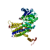


 PDBj
PDBj


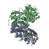
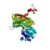
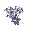
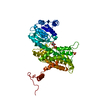
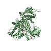
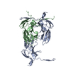
 Assembly
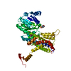


 Mass: 195.237 Da / Num. of mol.: 2 / Source method: obtained synthetically / Formula: C6H13NO4S / Comment: pH buffer*YM
 Mass: 18.015 Da / Num. of mol.: 580 / Source method: isolated from a natural source / Formula: H2O
 Sample preparation
 / Beamline: 24-ID-E / Wavelength: 0.97857 Å
 Processing