 Movie
Movie Controller

[English] 日本語
 Yorodumi
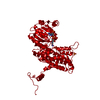
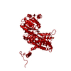
- PDB-1pgq: CRYSTALLOGRAPHIC STUDY OF COENZYME, COENZYME ANALOGUE AND SUBSTRA... -
+ Open data

- Basic information
| Entry | Database: PDB / ID: 1pgq | ||||||
|---|---|---|---|---|---|---|---|
| Title | CRYSTALLOGRAPHIC STUDY OF COENZYME, COENZYME ANALOGUE AND SUBSTRATE BINDING IN 6-PHOSPHOGLUCONATE DEHYDROGENASE: IMPLICATIONS FOR NADP SPECIFICITY AND THE ENZYME MECHANISM | ||||||
 Components Components | 6-PHOSPHOGLUCONATE DEHYDROGENASE | ||||||
 Keywords Keywords | OXIDOREDUCTASE (CHOH(D)-NADP+(A)) | ||||||
| Function / homology |  Function and homology information Function and homology informationD-gluconate metabolic process / phosphogluconate dehydrogenase (NADP+-dependent, decarboxylating) / phosphogluconate dehydrogenase (decarboxylating) activity / pentose-phosphate shunt / NADP binding / cytoplasmSimilarity search - Function | ||||||
| Biological species |  Ovis aries (sheep) Ovis aries (sheep) | ||||||
| Method | X-RAY DIFFRACTION / Resolution: 3.17 Å | ||||||
 Authors Authors | Adams, M.J. / Phillips, C. / Gover, S. | ||||||
 Citation Citation | Journal: Structure / Year: 1994 Title: Crystallographic study of coenzyme, coenzyme analogue and substrate binding in 6-phosphogluconate dehydrogenase: implications for NADP specificity and the enzyme mechanism. Authors: Adams, M.J. / Ellis, G.H. / Gover, S. / Naylor, C.E. / Phillips, C. #1: Journal: To be PublishedTitle: The Structure of 6-Phosphogluconate Dehydrogenase Refined at 2 Angstroms Resolution Authors: Phillips, C. / Gover, S. / Adams, M.J. #2: Journal: Acta Crystallogr.,Sect.B / Year: 1991Title: The Structure of 6-Phosphogluconate Dehydrogenase Refined at 2.5 Angstroms Resolution Authors: Adams, M.J. / Gover, S. / Leaback, R. / Phillips, C. / Somers, D.O'N. | ||||||
| History |
|
- Structure visualization
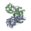
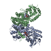
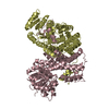
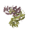
| Structure viewer | Molecule: MolmilJmol/JSmol |
|---|
- Downloads & links
-Download
| PDBx/mmCIF format | 1pgq.cif.gz | 112.7 KB | Display | PDBx/mmCIF format |
|---|---|---|---|---|
| PDB format | pdb1pgq.ent.gz | 86.2 KB | Display | PDB format |
| PDBx/mmJSON format | 1pgq.json.gz | Tree view | PDBx/mmJSON format | |
| Others |  Other downloads Other downloads |
-Validation report
| Arichive directory | https://data.pdbj.org/pub/pdb/validation_reports/pg/1pgqftp://data.pdbj.org/pub/pdb/validation_reports/pg/1pgq | HTTPS FTP |
|---|
-Related structure data


-Links
 PDBj
PDBj


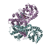
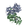
- Assembly
| Deposited unit | 
| ||||||||
|---|---|---|---|---|---|---|---|---|---|
| 1 | 
| ||||||||
| Unit cell |
|
-Components
| #1: Protein | Mass: 52905.652 Da / Num. of mol.: 1 Source method: isolated from a genetically manipulated source Source: (gene. exp.) Ovis aries (sheep)References: UniProt: P00349, phosphogluconate dehydrogenase (NADP+-dependent, decarboxylating) | ||||||
|---|---|---|---|---|---|---|---|
| #2: Chemical | Sulfate  Mass: 96.063 Da / Num. of mol.: 3 / Source method: obtained synthetically / Formula: SO4 Mass: 96.063 Da / Num. of mol.: 3 / Source method: obtained synthetically / Formula: SO4#3: Chemical | ChemComp-2AM / | Adenosine monophosphate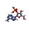  Mass: 347.221 Da / Num. of mol.: 1 / Source method: obtained synthetically / Formula: C10H14N5O7P Mass: 347.221 Da / Num. of mol.: 1 / Source method: obtained synthetically / Formula: C10H14N5O7P#4: Water | ChemComp-HOH / | Water Mass: 18.015 Da / Num. of mol.: 421 / Source method: isolated from a natural source / Formula: H2O Mass: 18.015 Da / Num. of mol.: 421 / Source method: isolated from a natural source / Formula: H2ONonpolymer details | THE HET GROUP 2AM, 2'-ADENYLIC ACID, CARRIES A -- CHARGE. | |
-Experimental details
-Experiment
| Experiment | Method: X-RAY DIFFRACTION |
|---|
- Sample preparation
| Crystal | Density Matthews: 2.61 Å3/Da / Density % sol: 52.87 % | |||||||||||||||
|---|---|---|---|---|---|---|---|---|---|---|---|---|---|---|---|---|
| Crystal grow | *PLUS pH: 6.5 / Method: batch method | |||||||||||||||
| Components of the solutions | *PLUS
|
-Data collection
| Radiation | Scattering type: x-ray |
|---|---|
| Radiation wavelength | Relative weight: 1 |
| Reflection | *PLUS Highest resolution: 3.17 Å / Num. obs: 8942 / % possible obs: 92.2 % / Num. measured all: 29208 / Rmerge(I) obs: 0.063 |
- Processing
| Software |
| ||||||||||||||||||||||||||||||||||||||||||||||||||||||||||||
|---|---|---|---|---|---|---|---|---|---|---|---|---|---|---|---|---|---|---|---|---|---|---|---|---|---|---|---|---|---|---|---|---|---|---|---|---|---|---|---|---|---|---|---|---|---|---|---|---|---|---|---|---|---|---|---|---|---|---|---|---|---|
| Refinement | Rfactor Rwork: 0.169 / Rfactor obs: 0.169 / Highest resolution: 3.17 Å Details: HET GROUP 2AM IS DESIGNATED 2'AMP IN THE JRNL REFERENCE. ATOMIC TEMPERATURE FACTORS AND SOLVENT OCCUPANCY AND POSITIONS WERE TAKEN FROM THE NATIVE STRUCTURE AT AN INTERMEDIATE STAGE OF ...Details: HET GROUP 2AM IS DESIGNATED 2'AMP IN THE JRNL REFERENCE. ATOMIC TEMPERATURE FACTORS AND SOLVENT OCCUPANCY AND POSITIONS WERE TAKEN FROM THE NATIVE STRUCTURE AT AN INTERMEDIATE STAGE OF REFINEMENT AND NOT REFINED. SOLVENT MOLECULES ARE ASSOCIATED WITH THE SAME MOLECULE AS THEIR CLOSEST OXYGEN OR NITROGEN NEIGHBOR; THIS BEST PRESERVES NETWORKS, BUT IN SOME INSTANCES HAS RESULTED IN THE NEAREST PROTEIN RESIDUE BEING THAT IN A SYMMETRY-RELATED MOLECULE. FOR SOME SOLVENT MOLECULES, THE DISTANCE TO THE CLOSEST OXYGEN OR NITROGEN NEIGHBOR IS MORE THAN 4 ANGSTROMS; THIS IS BECAUSE POORLY DEFINED SOLVENT SITES WERE OMITTED FROM NATIVE STRUCTURE REFINEMENT. THE CRITERIA APPLIED ARE DESCRIBED IN THE JRNL REFERENCE AND IN REFERENCE 1. | ||||||||||||||||||||||||||||||||||||||||||||||||||||||||||||
| Refinement step | Cycle: LAST / Highest resolution: 3.17 Å
| ||||||||||||||||||||||||||||||||||||||||||||||||||||||||||||
| Refine LS restraints |
| ||||||||||||||||||||||||||||||||||||||||||||||||||||||||||||
| Refinement | *PLUS Lowest resolution: 20 Å | ||||||||||||||||||||||||||||||||||||||||||||||||||||||||||||
| Solvent computation | *PLUS | ||||||||||||||||||||||||||||||||||||||||||||||||||||||||||||
| Displacement parameters | *PLUS | ||||||||||||||||||||||||||||||||||||||||||||||||||||||||||||
| Refine LS restraints | *PLUS
|
