 Movie
Movie Controller

[English] 日本語
 Yorodumi
- PDB-2pey: Crystal structure of deletion mutant of APS-kinase domain of huma... -
+ Open data

- Basic information
| Entry | Database: PDB / ID: 2pey | ||||||
|---|---|---|---|---|---|---|---|
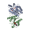
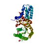
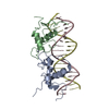
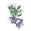
| Title | Crystal structure of deletion mutant of APS-kinase domain of human PAPS-synthetase 1 | ||||||
 Components Components | Bifunctional 3'-phosphoadenosine 5'-phosphosulfate synthetase 1 (PAPS synthetase 1) (PAPSS 1) (Sulfurylase kinase 1) (SK1) (SK 1) | ||||||
 Keywords Keywords |  TRANSFERASE / protein-nucleic acid complex / NMP-kinase fold TRANSFERASE / protein-nucleic acid complex / NMP-kinase fold | ||||||
| Function / homology |  Function and homology information Function and homology information3'-phosphoadenosine 5'-phosphosulfate biosynthetic process / Transport and synthesis of PAPS / adenylyl-sulfate kinase / sulfate adenylyltransferase / adenylylsulfate kinase activity / sulfate adenylyltransferase (ATP) activity / Metabolism of ingested H2SeO4 and H2SeO3 into H2Se / sulfate assimilation / nucleotidyltransferase activity / skeletal system development ...3'-phosphoadenosine 5'-phosphosulfate biosynthetic process / Transport and synthesis of PAPS / adenylyl-sulfate kinase / sulfate adenylyltransferase / adenylylsulfate kinase activity / sulfate adenylyltransferase (ATP) activity / Metabolism of ingested H2SeO4 and H2SeO3 into H2Se / sulfate assimilation / nucleotidyltransferase activity / skeletal system development / Signaling by BRAF and RAF1 fusions / phosphorylation / protein homodimerization activity / ATP binding / cytosolSimilarity search - Function | ||||||
| Biological species |  Homo sapiens (human) Homo sapiens (human) | ||||||
| Method | X-RAY DIFFRACTION / SYNCHROTRON / MOLECULAR REPLACEMENT / Resolution: 1.88 Å | ||||||
 Authors Authors | Sekulic, N. / Lavie, A. | ||||||
 Citation Citation | Journal: J.Biol.Chem. / Year: 2007 Title: Structural mechanism for substrate inhibition of the adenosine 5'-phosphosulfate kinase domain of human 3'-phosphoadenosine 5'-phosphosulfate synthetase 1 and its ramifications for enzyme regulation. Authors: Sekulic, N. / Konrad, M. / Lavie, A. | ||||||
| History |
|
- Structure visualization
| Structure viewer | Molecule: MolmilJmol/JSmol |
|---|
- Downloads & links
-Download
| PDBx/mmCIF format | 2pey.cif.gz | 83.9 KB | Display | PDBx/mmCIF format |
|---|---|---|---|---|
| PDB format | pdb2pey.ent.gz | 61.5 KB | Display | PDB format |
| PDBx/mmJSON format | 2pey.json.gz | Tree view | PDBx/mmJSON format | |
| Others |  Other downloads Other downloads |
-Validation report
| Arichive directory | https://data.pdbj.org/pub/pdb/validation_reports/pe/2peyftp://data.pdbj.org/pub/pdb/validation_reports/pe/2pey | HTTPS FTP |
|---|
-Related structure data
| Related structure data |  2pezC  2ofxS C: citing same article ( S: Starting model for refinement |
|---|---|
| Similar structure data |


-Links
 PDBj
PDBj
- Assembly
| Deposited unit | 
| ||||||||
|---|---|---|---|---|---|---|---|---|---|
| 1 |
| ||||||||
| Unit cell |
|
-Components
| #1: Protein | Mass: 19874.469 Da / Num. of mol.: 2 / Fragment: APS-kinase domain (residues 51-226) Source method: isolated from a genetically manipulated source Source: (gene. exp.) Homo sapiens (human) / Gene: PAPSS1, ATPSK1, PAPSSPlasmid: pGEX-RB in which the thrombin site was replaced by TEV cutting site Production host:  Escherichia coli (E. coli) / Strain (production host): BL-21 codon plus / References: UniProt: O43252, adenylyl-sulfate kinase Escherichia coli (E. coli) / Strain (production host): BL-21 codon plus / References: UniProt: O43252, adenylyl-sulfate kinase#2: Chemical | ChemComp-ADX / | 3'-Phosphoadenosine-5'-phosphosulfate  Type: RNA linking / Mass: 427.284 Da / Num. of mol.: 1 / Source method: obtained synthetically / Formula: C10H14N5O10PS Type: RNA linking / Mass: 427.284 Da / Num. of mol.: 1 / Source method: obtained synthetically / Formula: C10H14N5O10PS#3: Chemical | Deoxyadenosine diphosphate  Mass: 411.202 Da / Num. of mol.: 2 / Source method: obtained synthetically / Formula: C10H15N5O9P2 Mass: 411.202 Da / Num. of mol.: 2 / Source method: obtained synthetically / Formula: C10H15N5O9P2#4: Water | ChemComp-HOH / | Water Mass: 18.015 Da / Num. of mol.: 172 / Source method: isolated from a natural source / Formula: H2O Mass: 18.015 Da / Num. of mol.: 172 / Source method: isolated from a natural source / Formula: H2O |
|---|
-Experimental details
-Experiment
| Experiment | Method: X-RAY DIFFRACTION / Number of used crystals: 1 |
|---|
- Sample preparation
| Crystal | Density Matthews: 2.3 Å3/Da / Density % sol: 46.62 % |
|---|---|
| Crystal grow | Temperature: 293 K / Method: vapor diffusion, hanging drop / pH: 5 Details: reservoir: 16-20% PEG 3350, 0.25-0.15 M diammonium hydrogen citrate, drop: 3.2 mg/ml protein solution, 2mM dADP, 2mM APS, 5mM MgCl2, pH 5.0, VAPOR DIFFUSION, HANGING DROP, temperature 293K |
-Data collection
| Diffraction | Mean temperature: 193 K |
|---|---|
| Diffraction source | Source: SYNCHROTRON / Site: APS  / Beamline: 22-BM / Wavelength: 1 Å / Beamline: 22-BM / Wavelength: 1 Å |
| Detector | Type: MARMOSAIC 225 mm CCD / Detector: CCD / Date: Apr 7, 2006 |
| Radiation | Protocol: SINGLE WAVELENGTH / Monochromatic (M) / Laue (L): M / Scattering type: x-ray |
| Radiation wavelength | Wavelength: 1 Å / Relative weight: 1 |
| Reflection | Resolution: 1.88→20 Å / Num. all: 30062 / Num. obs: 29201 / % possible obs: 97 % / Observed criterion σ(F): 0 / Observed criterion σ(I): 0 / Redundancy: 6.9 % / Biso Wilson estimate: 24.6 Å2 / Rmerge(I) obs: 0.086 / Rsym value: 8.4 / Net I/σ(I): 16.63 |
| Reflection shell | Resolution: 1.88→2 Å / Redundancy: 5.4 % / Rmerge(I) obs: 0.388 / Mean I/σ(I) obs: 4.14 / Num. unique all: 3922 / Rsym value: 38.7 / % possible all: 82.6 |
- Processing
| Software |
| ||||||||||||||||||||||||||||||||||||||||||||||||||||||||||||||||||||||||||||||||||||||||||
|---|---|---|---|---|---|---|---|---|---|---|---|---|---|---|---|---|---|---|---|---|---|---|---|---|---|---|---|---|---|---|---|---|---|---|---|---|---|---|---|---|---|---|---|---|---|---|---|---|---|---|---|---|---|---|---|---|---|---|---|---|---|---|---|---|---|---|---|---|---|---|---|---|---|---|---|---|---|---|---|---|---|---|---|---|---|---|---|---|---|---|---|
| Refinement | Method to determine structure: MOLECULAR REPLACEMENT Starting model: PDB entry 2OFX Resolution: 1.88→20 Å / Cor.coef. Fo:Fc: 0.949 / Cor.coef. Fo:Fc free: 0.914 / SU B: 4.634 / SU ML: 0.13 / Cross valid method: THROUGHOUT / σ(F): 0 / σ(I): 0 / ESU R: 0.172 / ESU R Free: 0.165 / Stereochemistry target values: MAXIMUM LIKELIHOOD / Details: HYDROGENS HAVE BEEN ADDED IN THE RIDING POSITIONS
| ||||||||||||||||||||||||||||||||||||||||||||||||||||||||||||||||||||||||||||||||||||||||||
| Solvent computation | Ion probe radii: 0.8 Å / Shrinkage radii: 0.8 Å / VDW probe radii: 1.4 Å / Solvent model: MASK | ||||||||||||||||||||||||||||||||||||||||||||||||||||||||||||||||||||||||||||||||||||||||||
| Displacement parameters | Biso mean: 20.686 Å2
| ||||||||||||||||||||||||||||||||||||||||||||||||||||||||||||||||||||||||||||||||||||||||||
| Refinement step | Cycle: LAST / Resolution: 1.88→20 Å
| ||||||||||||||||||||||||||||||||||||||||||||||||||||||||||||||||||||||||||||||||||||||||||
| Refine LS restraints |
| ||||||||||||||||||||||||||||||||||||||||||||||||||||||||||||||||||||||||||||||||||||||||||
| LS refinement shell | Resolution: 1.88→1.933 Å / Total num. of bins used: 20
|
