 Movie
Movie Controller


+ Open data

- Basic information
| Entry | Database: PDB / ID: 2a18 | ||||||
|---|---|---|---|---|---|---|---|
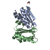
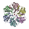
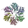
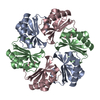
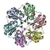
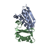
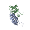
| Title | carboxysome shell protein ccmK4, crystal form 2 | ||||||
 Components Components | Carbon dioxide concentrating mechanism protein ccmK homolog 4 | ||||||
 Keywords Keywords |  carboxysome / cyclic hexamer / c6 point symmetry carboxysome / cyclic hexamer / c6 point symmetry | ||||||
| Function / homology |  Function and homology information Function and homology informationstructural constituent of carboxysome shell / carboxysome / carbon fixation / photosynthesisSimilarity search - Function | ||||||
| Biological species |  | ||||||
| Method | X-RAY DIFFRACTION / SYNCHROTRON / MOLECULAR REPLACEMENT / Resolution: 2.28 Å | ||||||
 Authors Authors | Kerfeld, C.A. / Sawaya, M.R. / Tanaka, S. / Nguyen, C.V. / Phillips, M. / Beeby, M. / Yeates, T.O. | ||||||
 Citation Citation | Journal: Science / Year: 2005 Title: Protein structures forming the shell of primitive bacterial organelles Authors: Kerfeld, C.A. / Sawaya, M.R. / Tanaka, S. / Nguyen, C.V. / Phillips, M. / Beeby, M. / Yeates, T.O. | ||||||
| History |
|
- Structure visualization
| Structure viewer | Molecule: MolmilJmol/JSmol |
|---|
- Downloads & links
-Download
| PDBx/mmCIF format | 2a18.cif.gz | 72.3 KB | Display | PDBx/mmCIF format |
|---|---|---|---|---|
| PDB format | pdb2a18.ent.gz | 53.6 KB | Display | PDB format |
| PDBx/mmJSON format | 2a18.json.gz | Tree view | PDBx/mmJSON format | |
| Others |  Other downloads Other downloads |
-Validation report
| Arichive directory | https://data.pdbj.org/pub/pdb/validation_reports/a1/2a18ftp://data.pdbj.org/pub/pdb/validation_reports/a1/2a18 | HTTPS FTP |
|---|
-Related structure data
| Related structure data | 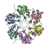 2a10SC 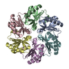 2a1bC S: Starting model for refinement C: citing same article ( |
|---|---|
| Similar structure data |

-Links
 PDBj
PDBj


- Assembly
| Deposited unit | 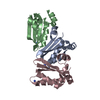
| ||||||||||||||||||||||||||||||||||||||||||||||||||||||||||||||||||||||||||||||||||||||||||||||||||||||||||||||||||||||||||||||||||||||||||||||||||||||||||||||||||||||||||||||||||||||||||
|---|---|---|---|---|---|---|---|---|---|---|---|---|---|---|---|---|---|---|---|---|---|---|---|---|---|---|---|---|---|---|---|---|---|---|---|---|---|---|---|---|---|---|---|---|---|---|---|---|---|---|---|---|---|---|---|---|---|---|---|---|---|---|---|---|---|---|---|---|---|---|---|---|---|---|---|---|---|---|---|---|---|---|---|---|---|---|---|---|---|---|---|---|---|---|---|---|---|---|---|---|---|---|---|---|---|---|---|---|---|---|---|---|---|---|---|---|---|---|---|---|---|---|---|---|---|---|---|---|---|---|---|---|---|---|---|---|---|---|---|---|---|---|---|---|---|---|---|---|---|---|---|---|---|---|---|---|---|---|---|---|---|---|---|---|---|---|---|---|---|---|---|---|---|---|---|---|---|---|---|---|---|---|---|---|---|---|---|
| 1 | 
| ||||||||||||||||||||||||||||||||||||||||||||||||||||||||||||||||||||||||||||||||||||||||||||||||||||||||||||||||||||||||||||||||||||||||||||||||||||||||||||||||||||||||||||||||||||||||||
| Unit cell |
| ||||||||||||||||||||||||||||||||||||||||||||||||||||||||||||||||||||||||||||||||||||||||||||||||||||||||||||||||||||||||||||||||||||||||||||||||||||||||||||||||||||||||||||||||||||||||||
| Noncrystallographic symmetry (NCS) | NCS domain:
NCS domain segments: Component-ID: 1 / Refine code: 1
|