 Movie
Movie Controller

[English] 日本語
 Yorodumi
- PDB-2oz5: Crystal structure of Mycobacterium tuberculosis protein tyrosine ... -
+ Open data

- Basic information
| Entry | Database: PDB / ID: 2oz5 | ||||||
|---|---|---|---|---|---|---|---|
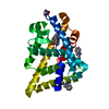
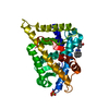
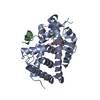
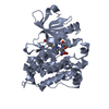
| Title | Crystal structure of Mycobacterium tuberculosis protein tyrosine phosphatase PtpB in complex with the specific inhibitor OMTS | ||||||
 Components Components | Phosphotyrosine protein phosphatase ptpb Tyrosine Tyrosine | ||||||
 Keywords Keywords | STRUCTURAL GENOMICS / UNKNOWN FUNCTION / Protein tyrosine phosphatase in complex with small molecule inhibitor / TB Structural Genomics Consortium / TBSGC | ||||||
| Function / homology |  Function and homology information Function and homology informationphosphatidylinositol trisphosphate phosphatase activity / biological process involved in interaction with host / protein serine/threonine phosphatase activity / phosphatase activity / protein dephosphorylation / protein tyrosine phosphatase activity / extracellular regionSimilarity search - Function | ||||||
| Biological species |   Mycobacterium tuberculosis (bacteria) Mycobacterium tuberculosis (bacteria) | ||||||
| Method | X-RAY DIFFRACTION / SYNCHROTRON / MOLECULAR REPLACEMENT / Resolution: 2 Å | ||||||
 Authors Authors | Grundner, C. / Gee, C.L. / Alber, T. / TB Structural Genomics Consortium (TBSGC) | ||||||
 Citation Citation | Journal: Structure / Year: 2007 Title: Structural Basis for Selective Inhibition of Mycobacterium tuberculosis Protein Tyrosine Phosphatase PtpB. Authors: Grundner, C. / Perrin, D. / Hooft van Huijsduijnen, R. / Swinnen, D. / Gonzalez, J. / Gee, C.L. / Wells, T.N. / Alber, T. | ||||||
| History |
|
- Structure visualization
| Structure viewer | Molecule: MolmilJmol/JSmol |
|---|
- Downloads & links
-Download
| PDBx/mmCIF format | 2oz5.cif.gz | 127.8 KB | Display | PDBx/mmCIF format |
|---|---|---|---|---|
| PDB format | pdb2oz5.ent.gz | 98.5 KB | Display | PDB format |
| PDBx/mmJSON format | 2oz5.json.gz | Tree view | PDBx/mmJSON format | |
| Others |  Other downloads Other downloads |
-Validation report
| Arichive directory | https://data.pdbj.org/pub/pdb/validation_reports/oz/2oz5ftp://data.pdbj.org/pub/pdb/validation_reports/oz/2oz5 | HTTPS FTP |
|---|
-Related structure data
| Similar structure data |
|---|


-Links
 PDBj
PDBj


- Assembly
| Deposited unit | 
| ||||||||
|---|---|---|---|---|---|---|---|---|---|
| 1 | 
| ||||||||
| 2 | 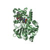
| ||||||||
| Unit cell |
|
-Components
| #1: Protein | Tyrosine Mass: 32365.191 Da / Num. of mol.: 2 Source method: isolated from a genetically manipulated source Source: (gene. exp.) Mycobacterium tuberculosis (bacteria) / Strain: H37Rv / Gene: ptpB / Plasmid: pET28b / Production host: Escherichia coli (E. coli) / Strain (production host): BL21 codon plus / References: UniProt: A1QMT0, UniProt: P96830*PLUS#2: Chemical | ChemComp-7XY / {(   Mass: 583.118 Da / Num. of mol.: 4 / Source method: obtained synthetically / Formula: C29H27ClN2O5S2 Mass: 583.118 Da / Num. of mol.: 4 / Source method: obtained synthetically / Formula: C29H27ClN2O5S2#3: Water | ChemComp-HOH / | Water Mass: 18.015 Da / Num. of mol.: 360 / Source method: isolated from a natural source / Formula: H2O Mass: 18.015 Da / Num. of mol.: 360 / Source method: isolated from a natural source / Formula: H2O |
|---|
-Experimental details
-Experiment
| Experiment | Method: X-RAY DIFFRACTION / Number of used crystals: 1 |
|---|
- Sample preparation
| Crystal | Density Matthews: 2 Å3/Da / Density % sol: 38.5 % |
|---|---|
| Crystal grow | Temperature: 291 K / Method: vapor diffusion, sitting drop / pH: 5.5 Details: 0.1M Ammonium acetate, 0.1M Bis-Tris pH5.5 17% PEG 10,000 0.5% beta-octylgucoside, VAPOR DIFFUSION, SITTING DROP, temperature 291K |
-Data collection
| Diffraction | Mean temperature: 100 K | |||||||||||||||||||||||||||||||||||||||||||||||||||||||||||||||||||||||||||||
|---|---|---|---|---|---|---|---|---|---|---|---|---|---|---|---|---|---|---|---|---|---|---|---|---|---|---|---|---|---|---|---|---|---|---|---|---|---|---|---|---|---|---|---|---|---|---|---|---|---|---|---|---|---|---|---|---|---|---|---|---|---|---|---|---|---|---|---|---|---|---|---|---|---|---|---|---|---|---|
| Diffraction source | Source: SYNCHROTRON / Site: ALS  / Beamline: 8.3.1 / Wavelength: 1.1159 Å / Beamline: 8.3.1 / Wavelength: 1.1159 Å | |||||||||||||||||||||||||||||||||||||||||||||||||||||||||||||||||||||||||||||
| Detector | Type: ADSC QUANTUM 4 / Detector: CCD / Date: Oct 6, 2006 | |||||||||||||||||||||||||||||||||||||||||||||||||||||||||||||||||||||||||||||
| Radiation | Monochromator: Si 111 channel / Protocol: SINGLE WAVELENGTH / Monochromatic (M) / Laue (L): M / Scattering type: x-ray | |||||||||||||||||||||||||||||||||||||||||||||||||||||||||||||||||||||||||||||
| Radiation wavelength | Wavelength: 1.1159 Å / Relative weight: 1 | |||||||||||||||||||||||||||||||||||||||||||||||||||||||||||||||||||||||||||||
| Reflection | Resolution: 2→50 Å / Num. obs: 34740 / % possible obs: 97.1 % / Redundancy: 3.6 % / Rmerge(I) obs: 0.034 / Χ2: 0.814 / Net I/σ(I): 16.5 | |||||||||||||||||||||||||||||||||||||||||||||||||||||||||||||||||||||||||||||
| Reflection shell |
|
-Phasing
| Phasing MR | Rfactor: 0.46 / Cor.coef. Fo:Fc: 0.472
|
|---|
- Processing
| Software |
| ||||||||||||||||||||||||||||||||||||||||||||||||||||||||||||||||||||||||||||||||||||||||||
|---|---|---|---|---|---|---|---|---|---|---|---|---|---|---|---|---|---|---|---|---|---|---|---|---|---|---|---|---|---|---|---|---|---|---|---|---|---|---|---|---|---|---|---|---|---|---|---|---|---|---|---|---|---|---|---|---|---|---|---|---|---|---|---|---|---|---|---|---|---|---|---|---|---|---|---|---|---|---|---|---|---|---|---|---|---|---|---|---|---|---|---|
| Refinement | Method to determine structure: MOLECULAR REPLACEMENT / Resolution: 2→50 Å / Cor.coef. Fo:Fc: 0.934 / Cor.coef. Fo:Fc free: 0.897 / SU B: 3.98 / SU ML: 0.116 / Cross valid method: THROUGHOUT / σ(F): 0 / ESU R: 0.212 / ESU R Free: 0.189 / Stereochemistry target values: MAXIMUM LIKELIHOOD Details: HYDROGENS HAVE BEEN ADDED IN THE RIDING POSITIONS. Some atoms in ARG 235 A, ARG 269 A, ARG 124 B, and ARG 258 B were modeled with zero occupancy leading to high RMS deviation values in these residues.
| ||||||||||||||||||||||||||||||||||||||||||||||||||||||||||||||||||||||||||||||||||||||||||
| Solvent computation | Ion probe radii: 0.8 Å / Shrinkage radii: 0.8 Å / VDW probe radii: 1.2 Å / Solvent model: MASK | ||||||||||||||||||||||||||||||||||||||||||||||||||||||||||||||||||||||||||||||||||||||||||
| Displacement parameters | Biso mean: 14.435 Å2
| ||||||||||||||||||||||||||||||||||||||||||||||||||||||||||||||||||||||||||||||||||||||||||
| Refinement step | Cycle: LAST / Resolution: 2→50 Å
| ||||||||||||||||||||||||||||||||||||||||||||||||||||||||||||||||||||||||||||||||||||||||||
| Refine LS restraints |
| ||||||||||||||||||||||||||||||||||||||||||||||||||||||||||||||||||||||||||||||||||||||||||
| LS refinement shell | Resolution: 2→2.052 Å / Total num. of bins used: 20
|
