 Movie
Movie Controller

[English] 日本語
 Yorodumi
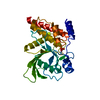
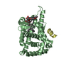
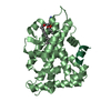
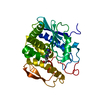
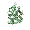
- PDB-1yuc: Human Nuclear Receptor Liver Receptor Homologue-1, LRH-1, Bound t... -
+ Open data

- Basic information
| Entry | Database: PDB / ID: 1yuc | ||||||
|---|---|---|---|---|---|---|---|
| Title | Human Nuclear Receptor Liver Receptor Homologue-1, LRH-1, Bound to Phospholipid and a Fragment of Human SHP | ||||||
 Components Components |
| ||||||
 Keywords Keywords |  TRANSCRIPTION REGULATION / Liver Receptor Homologue 1 / Nuclear Receptor Ligand Binding Domain / LRH-1 / Phospholipid / SHP / Small Heterodimer Partner TRANSCRIPTION REGULATION / Liver Receptor Homologue 1 / Nuclear Receptor Ligand Binding Domain / LRH-1 / Phospholipid / SHP / Small Heterodimer Partner | ||||||
| Function / homology |  Function and homology informationRegulation of gene expression in early pancreatic precursor cells / pancreas morphogenesis / calcineurin-mediated signaling / acinar cell differentiation / tissue development / bile acid metabolic process / bile acid and bile salt transport / transcription regulator inhibitor activity / peroxisome proliferator activated receptor binding / embryo development ending in birth or egg hatching ...Regulation of gene expression in early pancreatic precursor cells / pancreas morphogenesis / calcineurin-mediated signaling / acinar cell differentiation / tissue development / bile acid metabolic process / bile acid and bile salt transport / transcription regulator inhibitor activity / peroxisome proliferator activated receptor binding / embryo development ending in birth or egg hatching / nuclear thyroid hormone receptor binding / homeostatic process / animal organ regeneration / positive regulation of viral genome replication / response to glucose / nuclear retinoid X receptor binding / Notch signaling pathway / cholesterol metabolic process / hormone-mediated signaling pathway / cellular response to leukemia inhibitory factor / cholesterol homeostasis / transcription coregulator binding / circadian regulation of gene expression / SUMOylation of intracellular receptors / phospholipid binding / positive regulation of insulin secretion / negative regulation of DNA-binding transcription factor activity / response to organic cyclic compound / Nuclear Receptor transcription pathway / circadian rhythm / RNA polymerase II transcription regulator complex / transcription corepressor activity / nuclear receptor activity / sequence-specific double-stranded DNA binding / regulation of cell population proliferation / DNA-binding transcription activator activity, RNA polymerase II-specific / response to ethanol / Estrogen-dependent gene expression / sequence-specific DNA binding / transcription cis-regulatory region binding / DNA-binding transcription factor activity, RNA polymerase II-specific / RNA polymerase II cis-regulatory region sequence-specific DNA binding / DNA-binding transcription factor activity / protein domain specific binding / negative regulation of gene expression / intracellular membrane-bounded organelle / negative regulation of DNA-templated transcription / chromatin binding / chromatin / protein-containing complex binding / regulation of DNA-templated transcription / positive regulation of gene expression / regulation of transcription by RNA polymerase II / positive regulation of DNA-templated transcription / negative regulation of transcription by RNA polymerase II / protein homodimerization activity / positive regulation of transcription by RNA polymerase II / DNA binding / zinc ion binding / nucleoplasm / nucleus / cytoplasm Function and homology informationRegulation of gene expression in early pancreatic precursor cells / pancreas morphogenesis / calcineurin-mediated signaling / acinar cell differentiation / tissue development / bile acid metabolic process / bile acid and bile salt transport / transcription regulator inhibitor activity / peroxisome proliferator activated receptor binding / embryo development ending in birth or egg hatching ...Regulation of gene expression in early pancreatic precursor cells / pancreas morphogenesis / calcineurin-mediated signaling / acinar cell differentiation / tissue development / bile acid metabolic process / bile acid and bile salt transport / transcription regulator inhibitor activity / peroxisome proliferator activated receptor binding / embryo development ending in birth or egg hatching / nuclear thyroid hormone receptor binding / homeostatic process / animal organ regeneration / positive regulation of viral genome replication / response to glucose / nuclear retinoid X receptor binding / Notch signaling pathway / cholesterol metabolic process / hormone-mediated signaling pathway / cellular response to leukemia inhibitory factor / cholesterol homeostasis / transcription coregulator binding / circadian regulation of gene expression / SUMOylation of intracellular receptors / phospholipid binding / positive regulation of insulin secretion / negative regulation of DNA-binding transcription factor activity / response to organic cyclic compound / Nuclear Receptor transcription pathway / circadian rhythm / RNA polymerase II transcription regulator complex / transcription corepressor activity / nuclear receptor activity / sequence-specific double-stranded DNA binding / regulation of cell population proliferation / DNA-binding transcription activator activity, RNA polymerase II-specific / response to ethanol / Estrogen-dependent gene expression / sequence-specific DNA binding / transcription cis-regulatory region binding / DNA-binding transcription factor activity, RNA polymerase II-specific / RNA polymerase II cis-regulatory region sequence-specific DNA binding / DNA-binding transcription factor activity / protein domain specific binding / negative regulation of gene expression / intracellular membrane-bounded organelle / negative regulation of DNA-templated transcription / chromatin binding / chromatin / protein-containing complex binding / regulation of DNA-templated transcription / positive regulation of gene expression / regulation of transcription by RNA polymerase II / positive regulation of DNA-templated transcription / negative regulation of transcription by RNA polymerase II / protein homodimerization activity / positive regulation of transcription by RNA polymerase II / DNA binding / zinc ion binding / nucleoplasm / nucleus / cytoplasmSimilarity search - Function | ||||||
| Biological species |  Homo sapiens (human) Homo sapiens (human) | ||||||
| Method | X-RAY DIFFRACTION / SYNCHROTRON / MOLECULAR REPLACEMENT / Resolution: 1.9 Å | ||||||
 Authors Authors | Ortlund, E.A. / Yoonkwang, L. / Solomon, I.H. / Hager, J.M. / Safi, R. / Choi, Y. / Guan, Z. / Tripathy, A. / Raetz, C.R.H. / McDonnell, D.P. ...Ortlund, E.A. / Yoonkwang, L. / Solomon, I.H. / Hager, J.M. / Safi, R. / Choi, Y. / Guan, Z. / Tripathy, A. / Raetz, C.R.H. / McDonnell, D.P. / Moore, D.D. / Redinbo, M.R. | ||||||
 Citation Citation | Journal: Nat.Struct.Mol.Biol. / Year: 2005 Title: Modulation of human nuclear receptor LRH-1 activity by phospholipids and SHP Authors: Ortlund, E.A. / Lee, Y. / Solomon, I.H. / Hager, J.M. / Safi, R. / Choi, Y. / Guan, Z. / Tripathy, A. / Raetz, C.R.H. / McDonnell, D.P. / Moore, D.D. / Redinbo, M.R. | ||||||
| History |
|
- Structure visualization
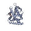
| Structure viewer | Molecule: MolmilJmol/JSmol |
|---|
- Downloads & links
-Download
| PDBx/mmCIF format | 1yuc.cif.gz | 120.6 KB | Display | PDBx/mmCIF format |
|---|---|---|---|---|
| PDB format | pdb1yuc.ent.gz | 94.1 KB | Display | PDB format |
| PDBx/mmJSON format | 1yuc.json.gz | Tree view | PDBx/mmJSON format | |
| Others |  Other downloads Other downloads |
-Validation report
| Arichive directory | https://data.pdbj.org/pub/pdb/validation_reports/yu/1yucftp://data.pdbj.org/pub/pdb/validation_reports/yu/1yuc | HTTPS FTP |
|---|
-Related structure data
| Similar structure data |
|---|


-Links
 PDBj
PDBj
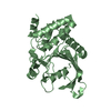
- Assembly
| Deposited unit | 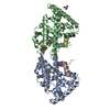
| ||||||||||
|---|---|---|---|---|---|---|---|---|---|---|---|
| 1 | 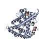
| ||||||||||
| 2 | 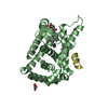
| ||||||||||
| 3 | 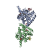
| ||||||||||
| 4 |
| ||||||||||
| Unit cell |
|
-Components
| #1: Protein | Mass: 29459.893 Da / Num. of mol.: 2 / Fragment: Ligand Binding Domain Source method: isolated from a genetically manipulated source Source: (gene. exp.) Homo sapiens (human) / Gene: NR5A2 / Plasmid: PMALCH10T / Production host:  Escherichia coli (E. coli) / References: UniProt: O00482 Escherichia coli (E. coli) / References: UniProt: O00482#2: Protein/peptide | Mass: 1449.673 Da / Num. of mol.: 2 / Fragment: NR Box1 / Source method: obtained synthetically / Details: sequence occurs natuarlly in Homo sapiens / References: UniProt: Q15466 #3: Chemical | Phosphatidylethanolamine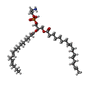  Mass: 709.933 Da / Num. of mol.: 2 / Source method: obtained synthetically / Formula: C39H68NO8P / Comment: phospholipid*YM Mass: 709.933 Da / Num. of mol.: 2 / Source method: obtained synthetically / Formula: C39H68NO8P / Comment: phospholipid*YM#4: Chemical | Glycerol  Mass: 92.094 Da / Num. of mol.: 3 / Source method: obtained synthetically / Formula: C3H8O3 Mass: 92.094 Da / Num. of mol.: 3 / Source method: obtained synthetically / Formula: C3H8O3#5: Water | ChemComp-HOH / | Water Mass: 18.015 Da / Num. of mol.: 245 / Source method: isolated from a natural source / Formula: H2O Mass: 18.015 Da / Num. of mol.: 245 / Source method: isolated from a natural source / Formula: H2O |
|---|
-Experimental details
-Experiment
| Experiment | Method: X-RAY DIFFRACTION / Number of used crystals: 1 |
|---|
- Sample preparation
| Crystal | Density Matthews: 2.3 Å3/Da / Density % sol: 46.5 % |
|---|---|
| Crystal grow | Method: vapor diffusion, hanging drop / pH: 6.4 Details: 9.5%-15% PEG 3350, 5% glycerol, and 50 mM Bis-Tris, pH 6.4, VAPOR DIFFUSION, HANGING DROP, temperature 100K |
-Data collection
| Diffraction | Mean temperature: 100 K |
|---|---|
| Diffraction source | Source: SYNCHROTRON / Site: SSRL  / Beamline: BL11-1 / Wavelength: 1 Å / Beamline: BL11-1 / Wavelength: 1 Å |
| Detector | Type: ADSC QUANTUM 315 / Detector: CCD / Date: May 15, 2004 |
| Radiation | Monochromator: Flat mirror (vertical focusing); single crystal Si(111) bent monochromator (horizontal focusing) Protocol: SINGLE WAVELENGTH / Monochromatic (M) / Laue (L): M / Scattering type: x-ray |
| Radiation wavelength | Wavelength: 1 Å / Relative weight: 1 |
| Reflection | Resolution: 1.9→50 Å / Num. obs: 42718 / % possible obs: 99.8 % / Observed criterion σ(I): 2 / Redundancy: 3.7 % / Biso Wilson estimate: 16.5 Å2 / Rsym value: 0.061 / Net I/σ(I): 22.6 |
| Reflection shell | Resolution: 1.9→1.97 Å / Redundancy: 3.7 % / Mean I/σ(I) obs: 4.15 / Rsym value: 0.309 / % possible all: 99.9 |
- Processing
| Software |
| |||||||||||||||||||||||||
|---|---|---|---|---|---|---|---|---|---|---|---|---|---|---|---|---|---|---|---|---|---|---|---|---|---|---|
| Refinement | Method to determine structure: MOLECULAR REPLACEMENT / Resolution: 1.9→31.28 Å / Rfactor Rfree error: 0.005 / Data cutoff high absF: 236131.39 / Data cutoff low absF: 0 / Isotropic thermal model: RESTRAINED / Cross valid method: THROUGHOUT / σ(F): 0 / Stereochemistry target values: Engh & Huber
| |||||||||||||||||||||||||
| Solvent computation | Solvent model: FLAT MODEL / Bsol: 45.6294 Å2 / ksol: 0.348664 e/Å3 | |||||||||||||||||||||||||
| Displacement parameters | Biso mean: 26.4 Å2
| |||||||||||||||||||||||||
| Refine analyze |
| |||||||||||||||||||||||||
| Refinement step | Cycle: LAST / Resolution: 1.9→31.28 Å
| |||||||||||||||||||||||||
| Refine LS restraints |
| |||||||||||||||||||||||||
| LS refinement shell | Resolution: 1.9→2.02 Å / Rfactor Rfree error: 0.015 / Total num. of bins used: 6
| |||||||||||||||||||||||||
| Xplor file |
|
