 Movie
Movie Controller

[English] 日本語
 Yorodumi
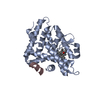
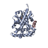
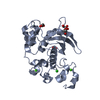
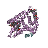
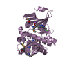
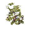
- PDB-4oni: Structure of Human Orphan Receptor LRH1 bound to two bacterial ph... -
+ Open data

- Basic information
| Entry | Database: PDB / ID: 4oni | ||||||
|---|---|---|---|---|---|---|---|
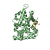
| Title | Structure of Human Orphan Receptor LRH1 bound to two bacterial phospholipids | ||||||
 Components Components | (Nuclear receptor subfamily ...) x 2 | ||||||
 Keywords Keywords |  NUCLEAR PROTEIN / alpha helical sandwich fold / Nuclear receptor / Co-factor binding NUCLEAR PROTEIN / alpha helical sandwich fold / Nuclear receptor / Co-factor binding | ||||||
| Function / homology |  Function and homology informationRegulation of gene expression in early pancreatic precursor cells / pancreas morphogenesis / calcineurin-mediated signaling / acinar cell differentiation / tissue development / bile acid metabolic process / bile acid and bile salt transport / transcription regulator inhibitor activity / peroxisome proliferator activated receptor binding / embryo development ending in birth or egg hatching ...Regulation of gene expression in early pancreatic precursor cells / pancreas morphogenesis / calcineurin-mediated signaling / acinar cell differentiation / tissue development / bile acid metabolic process / bile acid and bile salt transport / transcription regulator inhibitor activity / peroxisome proliferator activated receptor binding / embryo development ending in birth or egg hatching / nuclear thyroid hormone receptor binding / homeostatic process / animal organ regeneration / positive regulation of viral genome replication / response to glucose / nuclear retinoid X receptor binding / Notch signaling pathway / hormone-mediated signaling pathway / cholesterol metabolic process / cellular response to leukemia inhibitory factor / cholesterol homeostasis / transcription coregulator binding / phospholipid binding / circadian regulation of gene expression / SUMOylation of intracellular receptors / positive regulation of insulin secretion / negative regulation of DNA-binding transcription factor activity / response to organic cyclic compound / Nuclear Receptor transcription pathway / circadian rhythm / RNA polymerase II transcription regulator complex / transcription corepressor activity / nuclear receptor activity / sequence-specific double-stranded DNA binding / regulation of cell population proliferation / DNA-binding transcription activator activity, RNA polymerase II-specific / response to ethanol / Estrogen-dependent gene expression / sequence-specific DNA binding / transcription cis-regulatory region binding / DNA-binding transcription factor activity, RNA polymerase II-specific / RNA polymerase II cis-regulatory region sequence-specific DNA binding / DNA-binding transcription factor activity / protein domain specific binding / negative regulation of gene expression / intracellular membrane-bounded organelle / negative regulation of DNA-templated transcription / chromatin binding / chromatin / protein-containing complex binding / positive regulation of gene expression / regulation of DNA-templated transcription / regulation of transcription by RNA polymerase II / positive regulation of DNA-templated transcription / negative regulation of transcription by RNA polymerase II / protein homodimerization activity / positive regulation of transcription by RNA polymerase II / DNA binding / zinc ion binding / nucleoplasm / nucleus / cytoplasm Function and homology informationRegulation of gene expression in early pancreatic precursor cells / pancreas morphogenesis / calcineurin-mediated signaling / acinar cell differentiation / tissue development / bile acid metabolic process / bile acid and bile salt transport / transcription regulator inhibitor activity / peroxisome proliferator activated receptor binding / embryo development ending in birth or egg hatching ...Regulation of gene expression in early pancreatic precursor cells / pancreas morphogenesis / calcineurin-mediated signaling / acinar cell differentiation / tissue development / bile acid metabolic process / bile acid and bile salt transport / transcription regulator inhibitor activity / peroxisome proliferator activated receptor binding / embryo development ending in birth or egg hatching / nuclear thyroid hormone receptor binding / homeostatic process / animal organ regeneration / positive regulation of viral genome replication / response to glucose / nuclear retinoid X receptor binding / Notch signaling pathway / hormone-mediated signaling pathway / cholesterol metabolic process / cellular response to leukemia inhibitory factor / cholesterol homeostasis / transcription coregulator binding / phospholipid binding / circadian regulation of gene expression / SUMOylation of intracellular receptors / positive regulation of insulin secretion / negative regulation of DNA-binding transcription factor activity / response to organic cyclic compound / Nuclear Receptor transcription pathway / circadian rhythm / RNA polymerase II transcription regulator complex / transcription corepressor activity / nuclear receptor activity / sequence-specific double-stranded DNA binding / regulation of cell population proliferation / DNA-binding transcription activator activity, RNA polymerase II-specific / response to ethanol / Estrogen-dependent gene expression / sequence-specific DNA binding / transcription cis-regulatory region binding / DNA-binding transcription factor activity, RNA polymerase II-specific / RNA polymerase II cis-regulatory region sequence-specific DNA binding / DNA-binding transcription factor activity / protein domain specific binding / negative regulation of gene expression / intracellular membrane-bounded organelle / negative regulation of DNA-templated transcription / chromatin binding / chromatin / protein-containing complex binding / positive regulation of gene expression / regulation of DNA-templated transcription / regulation of transcription by RNA polymerase II / positive regulation of DNA-templated transcription / negative regulation of transcription by RNA polymerase II / protein homodimerization activity / positive regulation of transcription by RNA polymerase II / DNA binding / zinc ion binding / nucleoplasm / nucleus / cytoplasmSimilarity search - Function | ||||||
| Biological species |  Homo sapiens (human) Homo sapiens (human) | ||||||
| Method | X-RAY DIFFRACTION / SYNCHROTRON / MOLECULAR REPLACEMENT / molecular replacement / Resolution: 1.8 Å | ||||||
 Authors Authors | Gorman, M.A. / Parker, M.W. / Kusumo, S. | ||||||
 Citation Citation | Journal: To be Published Title: Human nuclear receptor LRH1 bound to phosopholipids and SHP peptide Authors: Gorman, M.A. / Parker, M.W. / Kusumo, S. | ||||||
| History |
|
- Structure visualization
| Structure viewer | Molecule: MolmilJmol/JSmol |
|---|
- Downloads & links
-Download
| PDBx/mmCIF format | 4oni.cif.gz | 133.6 KB | Display | PDBx/mmCIF format |
|---|---|---|---|---|
| PDB format | pdb4oni.ent.gz | 102.1 KB | Display | PDB format |
| PDBx/mmJSON format | 4oni.json.gz | Tree view | PDBx/mmJSON format | |
| Others |  Other downloads Other downloads |
-Validation report
| Arichive directory | https://data.pdbj.org/pub/pdb/validation_reports/on/4oniftp://data.pdbj.org/pub/pdb/validation_reports/on/4oni | HTTPS FTP |
|---|
-Related structure data
| Related structure data | 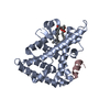 1yucS S: Starting model for refinement |
|---|---|
| Similar structure data |


-Links
 PDBj
PDBj
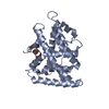
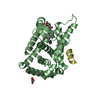
- Assembly
| Deposited unit | 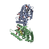
| ||||||||
|---|---|---|---|---|---|---|---|---|---|
| 1 | 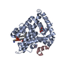
| ||||||||
| 2 | 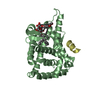
| ||||||||
| Unit cell |
|
-Components
-Nuclear receptor subfamily ... , 2 types, 4 molecules ABCD
| #1: Protein | Mass: 29196.541 Da / Num. of mol.: 2 / Fragment: Ligand Binding Domain, UNP residues 291-541 Source method: isolated from a genetically manipulated source Source: (gene. exp.) Homo sapiens (human) / Gene: NR5A2 / Plasmid: PMCSG7 / Production host:  Escherichia coli (E. coli) / Strain (production host): BL21 (DE3) Star Rosetta / References: UniProt: O00482 Escherichia coli (E. coli) / Strain (production host): BL21 (DE3) Star Rosetta / References: UniProt: O00482#2: Protein/peptide | Mass: 1948.269 Da / Num. of mol.: 2 / Fragment: NR Box1, UNP residues 12-30 / Source method: obtained synthetically / Details: This sequence occurs naturally in humans. / Source: (synth.) Homo sapiens (human) / References: UniProt: Q15466 |
|---|
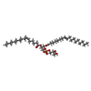
-Non-polymers , 5 types, 358 molecules 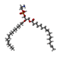


| #3: Chemical | ChemComp-EPH / Phosphatidylethanolamine Mass: 709.933 Da / Num. of mol.: 1 / Source method: obtained synthetically / Formula: C39H68NO8P / Comment: phospholipid*YM Mass: 709.933 Da / Num. of mol.: 1 / Source method: obtained synthetically / Formula: C39H68NO8P / Comment: phospholipid*YM | ||||||
|---|---|---|---|---|---|---|---|
| #4: Chemical | Glycerol Mass: 92.094 Da / Num. of mol.: 3 / Source method: obtained synthetically / Formula: C3H8O3 Mass: 92.094 Da / Num. of mol.: 3 / Source method: obtained synthetically / Formula: C3H8O3#5: Chemical | ChemComp-P6L / ( |  Mass: 746.991 Da / Num. of mol.: 1 / Source method: obtained synthetically / Formula: C40H75O10P / Comment: phospholipid*YM Mass: 746.991 Da / Num. of mol.: 1 / Source method: obtained synthetically / Formula: C40H75O10P / Comment: phospholipid*YM#6: Chemical | ChemComp-PG4 / | Polyethylene glycol Mass: 194.226 Da / Num. of mol.: 1 / Source method: obtained synthetically / Formula: C8H18O5 / Comment: precipitant*YM Mass: 194.226 Da / Num. of mol.: 1 / Source method: obtained synthetically / Formula: C8H18O5 / Comment: precipitant*YM#7: Water | ChemComp-HOH / | WaterMass: 18.015 Da / Num. of mol.: 352 / Source method: isolated from a natural source / Formula: H2O |
-Experimental details
-Experiment
| Experiment | Method: X-RAY DIFFRACTION / Number of used crystals: 1 |
|---|
- Sample preparation
| Crystal | Density Matthews: 2.26 Å3/Da / Density % sol: 45.67 % / Mosaicity: 0.16 ° |
|---|---|
| Crystal grow | Temperature: 298 K / Method: vapor diffusion, sitting drop / pH: 6.4 Details: 20-30% PEG 3350, Bis Tris, 5% glycerol, pH 6.4, VAPOR DIFFUSION, SITTING DROP, temperature 298K |
-Data collection
| Diffraction | Mean temperature: 100 K | ||||||||||||||||||||||||||||||||||||||||||||||||||||||||||||||||||||||||||||||||||||||||||||||||||||||||||||||||||||||||||||||||||||
|---|---|---|---|---|---|---|---|---|---|---|---|---|---|---|---|---|---|---|---|---|---|---|---|---|---|---|---|---|---|---|---|---|---|---|---|---|---|---|---|---|---|---|---|---|---|---|---|---|---|---|---|---|---|---|---|---|---|---|---|---|---|---|---|---|---|---|---|---|---|---|---|---|---|---|---|---|---|---|---|---|---|---|---|---|---|---|---|---|---|---|---|---|---|---|---|---|---|---|---|---|---|---|---|---|---|---|---|---|---|---|---|---|---|---|---|---|---|---|---|---|---|---|---|---|---|---|---|---|---|---|---|---|---|
| Diffraction source | Source: SYNCHROTRON / Site: Australian Synchrotron  / Beamline: MX2 / Wavelength: 0.96 Å / Beamline: MX2 / Wavelength: 0.96 Å | ||||||||||||||||||||||||||||||||||||||||||||||||||||||||||||||||||||||||||||||||||||||||||||||||||||||||||||||||||||||||||||||||||||
| Detector | Type: ADSC QUANTUM 315r / Detector: CCD / Date: Oct 3, 2013 | ||||||||||||||||||||||||||||||||||||||||||||||||||||||||||||||||||||||||||||||||||||||||||||||||||||||||||||||||||||||||||||||||||||
| Radiation | Monochromator: Si 111 CHANNEL / Protocol: SINGLE WAVELENGTH / Monochromatic (M) / Laue (L): M / Scattering type: x-ray | ||||||||||||||||||||||||||||||||||||||||||||||||||||||||||||||||||||||||||||||||||||||||||||||||||||||||||||||||||||||||||||||||||||
| Radiation wavelength | Wavelength: 0.96 Å / Relative weight: 1 | ||||||||||||||||||||||||||||||||||||||||||||||||||||||||||||||||||||||||||||||||||||||||||||||||||||||||||||||||||||||||||||||||||||
| Reflection | Resolution: 1.8→73.772 Å / Num. all: 49141 / Num. obs: 49141 / % possible obs: 94.8 % / Observed criterion σ(F): 0 / Observed criterion σ(I): 0 / Redundancy: 7 % / Rsym value: 0.119 / Net I/σ(I): 13.2 | ||||||||||||||||||||||||||||||||||||||||||||||||||||||||||||||||||||||||||||||||||||||||||||||||||||||||||||||||||||||||||||||||||||
| Reflection shell | Diffraction-ID: 1
|
-Phasing
| Phasing | Method: molecular replacement |
|---|
- Processing
| Software |
| ||||||||||||||||||||||||||||||||||||||||||||||||||||||||||||
|---|---|---|---|---|---|---|---|---|---|---|---|---|---|---|---|---|---|---|---|---|---|---|---|---|---|---|---|---|---|---|---|---|---|---|---|---|---|---|---|---|---|---|---|---|---|---|---|---|---|---|---|---|---|---|---|---|---|---|---|---|---|
| Refinement | Method to determine structure: MOLECULAR REPLACEMENT Starting model: 1YUC Resolution: 1.8→43.67 Å / Cor.coef. Fo:Fc: 0.962 / Cor.coef. Fo:Fc free: 0.938 / WRfactor Rfree: 0.1894 / WRfactor Rwork: 0.1458 / Occupancy max: 1 / Occupancy min: 0.5 / FOM work R set: 0.8936 / SU B: 2.416 / SU ML: 0.075 / SU R Cruickshank DPI: 0.1211 / SU Rfree: 0.1208 / Cross valid method: THROUGHOUT / σ(F): 0 / ESU R: 0.121 / ESU R Free: 0.121 / Stereochemistry target values: MAXIMUM LIKELIHOOD Details: HYDROGENS HAVE BEEN ADDED IN THE RIDING POSITIONS U VALUES: REFINED INDIVIDUALLY
| ||||||||||||||||||||||||||||||||||||||||||||||||||||||||||||
| Solvent computation | Ion probe radii: 0.8 Å / Shrinkage radii: 0.8 Å / VDW probe radii: 1.2 Å / Solvent model: MASK | ||||||||||||||||||||||||||||||||||||||||||||||||||||||||||||
| Displacement parameters | Biso max: 72.13 Å2 / Biso mean: 19.7988 Å2 / Biso min: 5.76 Å2
| ||||||||||||||||||||||||||||||||||||||||||||||||||||||||||||
| Refinement step | Cycle: LAST / Resolution: 1.8→43.67 Å
| ||||||||||||||||||||||||||||||||||||||||||||||||||||||||||||
| Refine LS restraints |
| ||||||||||||||||||||||||||||||||||||||||||||||||||||||||||||
| LS refinement shell | Resolution: 1.8→1.847 Å / Total num. of bins used: 20
|
