 Movie
Movie Controller


+ Open data

- Basic information
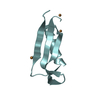
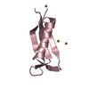
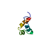
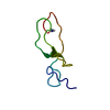
| Entry | Database: PDB / ID: 2op7 | ||||||
|---|---|---|---|---|---|---|---|
| Title | WW4 | ||||||
 Components Components | NEDD4-like E3 ubiquitin-protein ligase WWP1 | ||||||
 Keywords Keywords |  LIGASE / WW domain / Beta sheet LIGASE / WW domain / Beta sheet | ||||||
| Function / homology |  Function and homology information Function and homology informationHECT-type E3 ubiquitin transferase / ubiquitin ligase complex / Downregulation of ERBB4 signaling / monoatomic ion transmembrane transport / central nervous system development / Stimuli-sensing channels / ubiquitin-protein transferase activity / Regulation of RUNX2 expression and activity / ubiquitin protein ligase activity / Antigen processing: Ubiquitination & Proteasome degradation ...HECT-type E3 ubiquitin transferase / ubiquitin ligase complex / Downregulation of ERBB4 signaling / monoatomic ion transmembrane transport / central nervous system development / Stimuli-sensing channels / ubiquitin-protein transferase activity / Regulation of RUNX2 expression and activity / ubiquitin protein ligase activity / Antigen processing: Ubiquitination & Proteasome degradation / proteasome-mediated ubiquitin-dependent protein catabolic process / protein ubiquitination / symbiont entry into host cell / negative regulation of DNA-templated transcription / signal transduction / extracellular exosome / nucleus / plasma membrane / cytosol / cytoplasmSimilarity search - Function | ||||||
| Biological species |  Homo sapiens (human) Homo sapiens (human) | ||||||
| Method | SOLUTION NMR / distance geometry | ||||||
 Authors Authors | Qin, H.N. / Li, M.F. / Pu, H. / Sankaran, S. / Ahmed, S. / Song, J.X. | ||||||
 Citation Citation | Journal: To be Published Title: NMR structure of the forth WW domain of WWP1 Authors: Qin, H.N. / Li, M.F. / Pu, H. / Sankaran, S. / Ahmed, S. / Song, J.X. | ||||||
| History |
|
- Structure visualization
| Structure viewer | Molecule: MolmilJmol/JSmol |
|---|
- Downloads & links
-Download
| PDBx/mmCIF format | 2op7.cif.gz | 122.1 KB | Display | PDBx/mmCIF format |
|---|---|---|---|---|
| PDB format | pdb2op7.ent.gz | 98.4 KB | Display | PDB format |
| PDBx/mmJSON format | 2op7.json.gz | Tree view | PDBx/mmJSON format | |
| Others |  Other downloads Other downloads |
-Validation report
| Arichive directory | https://data.pdbj.org/pub/pdb/validation_reports/op/2op7ftp://data.pdbj.org/pub/pdb/validation_reports/op/2op7 | HTTPS FTP |
|---|
-Related structure data
| Similar structure data |
|---|


-Links
 PDBj
PDBj


- Assembly
| Deposited unit | 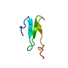
| |||||||||
|---|---|---|---|---|---|---|---|---|---|---|
| 1 |
| |||||||||
| NMR ensembles |
|
-Components
| #1: Protein/peptide | Mass: 4705.121 Da / Num. of mol.: 1 / Fragment: residues 494-532 Source method: isolated from a genetically manipulated source Source: (gene. exp.) Homo sapiens (human) / Gene: WWP1 / Plasmid: PGEX4T1 / Species (production host): Escherichia coli / Production host:  Escherichia coli BL21 (bacteria) / Strain (production host): BL21 Escherichia coli BL21 (bacteria) / Strain (production host): BL21References: UniProt: Q9H0M0, Ligases; Forming carbon-nitrogen bonds; Acid-amino-acid ligases (peptide synthases) |
|---|
-Experimental details
-Experiment
| Experiment | Method: SOLUTION NMR | ||||||||||||||||
|---|---|---|---|---|---|---|---|---|---|---|---|---|---|---|---|---|---|
| NMR experiment |
|
- Sample preparation
| Details | Solvent system: 90% H2O/10% D2O |
|---|---|
| Sample conditions | pH: 6.3 / Pressure: ambient / Temperature: 298 K |
-NMR measurement
| Radiation | Protocol: SINGLE WAVELENGTH / Monochromatic (M) / Laue (L): M |
|---|---|
| Radiation wavelength | Relative weight: 1 |
| NMR spectrometer | Type: Bruker INOVA / Manufacturer: Bruker / Model: INOVA / Field strength: 800 MHz |
- Processing
| NMR software |
| ||||||||||||
|---|---|---|---|---|---|---|---|---|---|---|---|---|---|
| Refinement | Method: distance geometry / Software ordinal: 1 | ||||||||||||
| NMR representative | Selection criteria: closest to the average | ||||||||||||
| NMR ensemble | Conformer selection criteria: target function / Conformers calculated total number: 1000 / Conformers submitted total number: 10 |
