 Movie
Movie Controller

[English] 日本語
 Yorodumi
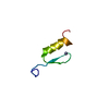
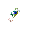
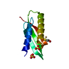
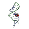
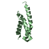
- PDB-1hz6: CRYSTAL STRUCTURES OF THE B1 DOMAIN OF PROTEIN L FROM PEPTOSTREPT... -
+ Open data

- Basic information
| Entry | Database: PDB / ID: 1hz6 | ||||||
|---|---|---|---|---|---|---|---|
| Title | CRYSTAL STRUCTURES OF THE B1 DOMAIN OF PROTEIN L FROM PEPTOSTREPTOCOCCUS MAGNUS WITH A TYROSINE TO TRYPTOPHAN SUBSTITUTION | ||||||
 Components Components | PROTEIN L | ||||||
 Keywords Keywords | PROTEIN BINDING / Four stranded beta-sheet with central alpha helix / binds kappa light chain of immunoglobulins | ||||||
| Function / homology |  Function and homology information Function and homology information | ||||||
| Biological species |  Finegoldia magna (bacteria) Finegoldia magna (bacteria) | ||||||
| Method | X-RAY DIFFRACTION / MOLECULAR REPLACEMENT / Resolution: 1.7 Å | ||||||
 Authors Authors | O'Neill, J.W. / Kim, D.E. / Baker, D. / Zhang, K.Y.J. | ||||||
 Citation Citation | Journal: Acta Crystallogr.,Sect.D / Year: 2001 Title: Structures of the B1 domain of protein L from Peptostreptococcus magnus with a tyrosine to tryptophan substitution. Authors: O'Neill, J.W. / Kim, D.E. / Baker, D. / Zhang, K.Y. | ||||||
| History |
|
- Structure visualization
| Structure viewer | Molecule: MolmilJmol/JSmol |
|---|
- Downloads & links
-Download
| PDBx/mmCIF format | 1hz6.cif.gz | 55 KB | Display | PDBx/mmCIF format |
|---|---|---|---|---|
| PDB format | pdb1hz6.ent.gz | 40 KB | Display | PDB format |
| PDBx/mmJSON format | 1hz6.json.gz | Tree view | PDBx/mmJSON format | |
| Others |  Other downloads Other downloads |
-Validation report
| Arichive directory | https://data.pdbj.org/pub/pdb/validation_reports/hz/1hz6ftp://data.pdbj.org/pub/pdb/validation_reports/hz/1hz6 | HTTPS FTP |
|---|
-Related structure data


-Links
 PDBj
PDBj- Assembly
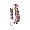
| Deposited unit | 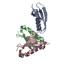
| ||||||||
|---|---|---|---|---|---|---|---|---|---|
| 1 | 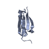
| ||||||||
| 2 | 
| ||||||||
| 3 | 
| ||||||||
| 4 |
| ||||||||
| 5 | 
| ||||||||
| Unit cell |
|
-Components
| #1: Protein | / IG KAPPA LIGHT CHAIN-BINDING PROTEIN Mass: 8052.908 Da / Num. of mol.: 3 / Fragment: B1 DOMAIN / Mutation: Y47W Source method: isolated from a genetically manipulated source Source: (gene. exp.) Finegoldia magna (bacteria) / Strain: ATCC 29328 / Plasmid: PET3A / Species (production host): Escherichia coli / Production host: Escherichia coli BL21(DE3) (bacteria) / Strain (production host): BL21(DE3) / References: UniProt: Q51912#2: Water | ChemComp-HOH / | Water Mass: 18.015 Da / Num. of mol.: 299 / Source method: isolated from a natural source / Formula: H2O Mass: 18.015 Da / Num. of mol.: 299 / Source method: isolated from a natural source / Formula: H2O |
|---|
-Experimental details
-Experiment
| Experiment | Method: X-RAY DIFFRACTION / Number of used crystals: 1 |
|---|
- Sample preparation
| Crystal | Density Matthews: 2.75 Å3/Da / Density % sol: 54.9 % | ||||||||||||||||||||||||
|---|---|---|---|---|---|---|---|---|---|---|---|---|---|---|---|---|---|---|---|---|---|---|---|---|---|
| Crystal grow | Temperature: 277 K / Method: vapor diffusion, hanging drop / pH: 5.5 Details: 30%PEG 8000, 0.2M Ammonium Sulfate, pH 5.5, VAPOR DIFFUSION, HANGING DROP, temperature 277K | ||||||||||||||||||||||||
| Crystal grow | *PLUS | ||||||||||||||||||||||||
| Components of the solutions | *PLUS
|
-Data collection
| Diffraction | Mean temperature: 100 K |
|---|---|
| Diffraction source | Source: ROTATING ANODE / Type: RIGAKU / Wavelength: 1.5418 Å |
| Detector | Type: RIGAKU RAXIS IV / Detector: IMAGE PLATE / Date: Oct 5, 1999 / Details: Mirrors |
| Radiation | Monochromator: YALE MIRRORS / Protocol: SINGLE WAVELENGTH / Monochromatic (M) / Laue (L): M / Scattering type: x-ray |
| Radiation wavelength | Wavelength: 1.5418 Å / Relative weight: 1 |
| Reflection | Resolution: 1.7→25 Å / Num. all: 120968 / Num. obs: 28825 / % possible obs: 96.3 % / Observed criterion σ(F): 0 / Observed criterion σ(I): -3 / Redundancy: 4.94 % / Biso Wilson estimate: 23.6 Å2 / Rmerge(I) obs: 0.074 / Rsym value: 0.08 / Net I/σ(I): 17 |
| Reflection shell | Resolution: 1.69→1.71 Å / Redundancy: 4.9 % / Rmerge(I) obs: 0.273 / Mean I/σ(I) obs: 6.8 / Num. unique all: 1031 / Rsym value: 0.252 / % possible all: 95.1 |
| Reflection shell | *PLUS % possible obs: 95.1 % |
- Processing
| Software |
| ||||||||||||||||||||||||||||||||||||
|---|---|---|---|---|---|---|---|---|---|---|---|---|---|---|---|---|---|---|---|---|---|---|---|---|---|---|---|---|---|---|---|---|---|---|---|---|---|
| Refinement | Method to determine structure: MOLECULAR REPLACEMENT Starting model: C3 DOMAIN OF PROTEIN L (UNPUBLISHED: T. WAN AND B. SUTTON) Resolution: 1.7→25 Å / Rfactor Rfree error: 0.006 / Data cutoff high absF: 923930.08 / Data cutoff low absF: 0 / Isotropic thermal model: RESTRAINED / Cross valid method: THROUGHOUT / σ(F): 0 / σ(I): -3 / Stereochemistry target values: MAXIMUM LIKELIHOOD
| ||||||||||||||||||||||||||||||||||||
| Solvent computation | Solvent model: FLAT MODEL / Bsol: 51.29 Å2 / ksol: 0.368 e/Å3 | ||||||||||||||||||||||||||||||||||||
| Displacement parameters | Biso mean: 24.2 Å2
| ||||||||||||||||||||||||||||||||||||
| Refine analyze |
| ||||||||||||||||||||||||||||||||||||
| Refinement step | Cycle: LAST / Resolution: 1.7→25 Å
| ||||||||||||||||||||||||||||||||||||
| Refine LS restraints |
| ||||||||||||||||||||||||||||||||||||
| LS refinement shell | Resolution: 1.7→1.76 Å / Rfactor Rfree error: 0.019 / Total num. of bins used: 6
| ||||||||||||||||||||||||||||||||||||
| Xplor file |
| ||||||||||||||||||||||||||||||||||||
| Software | *PLUS Name: CNS / Version: 1 / Classification: refinement | ||||||||||||||||||||||||||||||||||||
| Refine LS restraints | *PLUS
|
