 Movie
Movie Controller

[English] 日本語
 Yorodumi
- PDB-2odm: Crystal structure of S. aureus YlaN, an essential leucine rich pr... -
+ Open data

- Basic information
| Entry | Database: PDB / ID: 2odm | ||||||
|---|---|---|---|---|---|---|---|
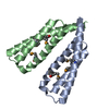
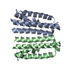
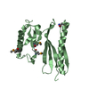
| Title | Crystal structure of S. aureus YlaN, an essential leucine rich protein involved in the control of cell shape | ||||||
 Components Components | UPF0358 protein MW0995 | ||||||
 Keywords Keywords | UNKNOWN FUNCTION /  triple helix triple helix | ||||||
| Function / homology | Uncharacterised protein family UPF0358 / Protein of unknown function (DUF1507) / SO2669-like / UPF0358 superfamily / Helix Hairpins / Orthogonal Bundle / Mainly Alpha / UPF0358 protein MW0995 Function and homology information Function and homology information | ||||||
| Biological species |   Staphylococcus aureus subsp. aureus (bacteria) Staphylococcus aureus subsp. aureus (bacteria) | ||||||
| Method | X-RAY DIFFRACTION / SYNCHROTRON / MAD / Resolution: 2.24 Å | ||||||
 Authors Authors | Xu, L. / Sedelnikova, S.E. / Baker, P.J. / Errington, J. / Hunt, A. / Rice, D.W. | ||||||
 Citation Citation | Journal: Proteins / Year: 2007 Title: Crystal structure of S. aureus YlaN, an essential leucine rich protein involved in the control of cell shape. Authors: Xu, L. / Sedelnikova, S.E. / Baker, P.J. / Hunt, A. / Errington, J. / Rice, D.W. | ||||||
| History |
|
- Structure visualization
| Structure viewer | Molecule: MolmilJmol/JSmol |
|---|
- Downloads & links
-Download
| PDBx/mmCIF format | 2odm.cif.gz | 43.1 KB | Display | PDBx/mmCIF format |
|---|---|---|---|---|
| PDB format | pdb2odm.ent.gz | 31.8 KB | Display | PDB format |
| PDBx/mmJSON format | 2odm.json.gz | Tree view | PDBx/mmJSON format | |
| Others |  Other downloads Other downloads |
-Validation report
| Arichive directory | https://data.pdbj.org/pub/pdb/validation_reports/od/2odmftp://data.pdbj.org/pub/pdb/validation_reports/od/2odm | HTTPS FTP |
|---|
-Related structure data
| Similar structure data |
|---|


-Links
 PDBj
PDBj- Assembly
| Deposited unit | 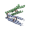
| ||||||||
|---|---|---|---|---|---|---|---|---|---|
| 1 |
| ||||||||
| Unit cell |
| ||||||||
| Details | biological dimer |
-Components
| #1: Protein | Mass: 10556.743 Da / Num. of mol.: 2 Source method: isolated from a genetically manipulated source Source: (gene. exp.) Staphylococcus aureus subsp. aureus (bacteria)Species: Staphylococcus aureus / Strain: MW2 / Gene: ylaN / Plasmid: pETBLUE1 / Production host: Escherichia coli (E. coli) / References: UniProt: Q7A161 |
|---|
-Experimental details
-Experiment
| Experiment | Method: X-RAY DIFFRACTION / Number of used crystals: 1 |
|---|
- Sample preparation
| Crystal | Density Matthews: 1.99 Å3/Da / Density % sol: 38.2 % |
|---|---|
| Crystal grow | Temperature: 291 K / Method: vapor diffusion, hanging drop / pH: 8 Details: 0.2M Sodium acetate, 0.1M Tris-HCl pH 8.5, 20% PEG4000, pH 8.0, VAPOR DIFFUSION, HANGING DROP, temperature 291K |
-Data collection
| Diffraction |
| |||||||||||||||
|---|---|---|---|---|---|---|---|---|---|---|---|---|---|---|---|---|
| Diffraction source |
| |||||||||||||||
| Detector | Type: MAR CCD 165 mm / Detector: CCD / Date: Jun 26, 2005 | |||||||||||||||
| Radiation | Monochromator: double crystal Si(III) / Protocol: MAD / Monochromatic (M) / Laue (L): M / Scattering type: x-ray | |||||||||||||||
| Radiation wavelength |
| |||||||||||||||
| Reflection | Resolution: 2.24→25 Å / Num. all: 7464 / % possible obs: 100 % / Observed criterion σ(F): 0 / Observed criterion σ(I): 0 / Redundancy: 3.6 % / Biso Wilson estimate: 51 Å2 / Rmerge(I) obs: 0.079 / Rsym value: 0.079 / Net I/σ(I): 10.5 | |||||||||||||||
| Reflection shell | Highest resolution: 2.24 Å / Redundancy: 3.6 % / Rmerge(I) obs: 0.429 / Mean I/σ(I) obs: 2.6 / Num. unique all: 1072 / Rsym value: 0.429 / % possible all: 99.7 |

- Processing
| Software |
| ||||||||||||||||||||||||||||||||||||||||||||||||||||||||||||||||||||||||||||||||||||||||||
|---|---|---|---|---|---|---|---|---|---|---|---|---|---|---|---|---|---|---|---|---|---|---|---|---|---|---|---|---|---|---|---|---|---|---|---|---|---|---|---|---|---|---|---|---|---|---|---|---|---|---|---|---|---|---|---|---|---|---|---|---|---|---|---|---|---|---|---|---|---|---|---|---|---|---|---|---|---|---|---|---|---|---|---|---|---|---|---|---|---|---|---|
| Refinement | Method to determine structure: MAD / Resolution: 2.24→15 Å / Cor.coef. Fo:Fc: 0.95 / Cor.coef. Fo:Fc free: 0.935 / SU B: 8.566 / SU ML: 0.214 / Isotropic thermal model: isotropic / Cross valid method: THROUGHOUT / σ(F): 0 / σ(I): 0 / ESU R: 0.369 / ESU R Free: 0.266 / Stereochemistry target values: MAXIMUM LIKELIHOOD
| ||||||||||||||||||||||||||||||||||||||||||||||||||||||||||||||||||||||||||||||||||||||||||
| Solvent computation | Ion probe radii: 0.8 Å / Shrinkage radii: 0.8 Å / VDW probe radii: 1.2 Å / Solvent model: MASK | ||||||||||||||||||||||||||||||||||||||||||||||||||||||||||||||||||||||||||||||||||||||||||
| Displacement parameters | Biso mean: 59.86 Å2
| ||||||||||||||||||||||||||||||||||||||||||||||||||||||||||||||||||||||||||||||||||||||||||
| Refinement step | Cycle: LAST / Resolution: 2.24→15 Å
| ||||||||||||||||||||||||||||||||||||||||||||||||||||||||||||||||||||||||||||||||||||||||||
| Refine LS restraints |
| ||||||||||||||||||||||||||||||||||||||||||||||||||||||||||||||||||||||||||||||||||||||||||
| LS refinement shell | Resolution: 2.24→2.296 Å / Total num. of bins used: 20
|
