 Movie
Movie Controller


+ Open data

- Basic information
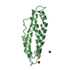
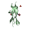
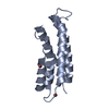
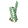
| Entry | Database: PDB / ID: 5wd8 | ||||||
|---|---|---|---|---|---|---|---|
| Title | Crystal structure of Legionella pneumophila effector lpg2328 | ||||||
 Components Components | Lem22 | ||||||
 Keywords Keywords |  PROTEIN BINDING / bacterial effector / Legionella / Structural Genomics / Montreal-Kingston Bacterial Structural Genomics Initiative / BSGI PROTEIN BINDING / bacterial effector / Legionella / Structural Genomics / Montreal-Kingston Bacterial Structural Genomics Initiative / BSGI | ||||||
| Function / homology | :  Function and homology information Function and homology information | ||||||
| Biological species |  Legionella pneumophila subsp. pneumophila ATCC 43290 (bacteria) Legionella pneumophila subsp. pneumophila ATCC 43290 (bacteria) | ||||||
| Method | X-RAY DIFFRACTION / SYNCHROTRON / SAD / Resolution: 1.944 Å | ||||||
 Authors Authors | Kozlov, G. / Wong, K. / Gehring, K. / Montreal-Kingston Bacterial Structural Genomics Initiative (BSGI) | ||||||
| Funding support |  Canada, 1items Canada, 1items
| ||||||
 Citation Citation | Journal: Proteins / Year: 2018 Title: Crystal structure of the Legionella effector Lem22. Authors: Kozlov, G. / Wong, K. / Gehring, K. | ||||||
| History |
|
- Structure visualization
| Structure viewer | Molecule: MolmilJmol/JSmol |
|---|
- Downloads & links
-Download
| PDBx/mmCIF format | 5wd8.cif.gz | 84.9 KB | Display | PDBx/mmCIF format |
|---|---|---|---|---|
| PDB format | pdb5wd8.ent.gz | 68.3 KB | Display | PDB format |
| PDBx/mmJSON format | 5wd8.json.gz | Tree view | PDBx/mmJSON format | |
| Others |  Other downloads Other downloads |
-Validation report
| Arichive directory | https://data.pdbj.org/pub/pdb/validation_reports/wd/5wd8ftp://data.pdbj.org/pub/pdb/validation_reports/wd/5wd8 | HTTPS FTP |
|---|
-Related structure data


-Links
 PDBj
PDBj- Assembly
| Deposited unit | 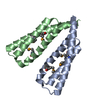
| ||||||||
|---|---|---|---|---|---|---|---|---|---|
| 1 | 
| ||||||||
| 2 | 
| ||||||||
| Unit cell |
|
-Components
| #1: Protein | Mass: 10670.622 Da / Num. of mol.: 2 Source method: isolated from a genetically manipulated source Source: (gene. exp.) Legionella pneumophila subsp. pneumophila ATCC 43290 (bacteria)Gene: lp12_2320 / Production host: Escherichia coli (E. coli) / Strain (production host): BL21 / References: UniProt: G8UTY6#2: Water | ChemComp-HOH / | Water Mass: 18.015 Da / Num. of mol.: 174 / Source method: isolated from a natural source / Formula: H2O Mass: 18.015 Da / Num. of mol.: 174 / Source method: isolated from a natural source / Formula: H2O |
|---|
-Experimental details
-Experiment
| Experiment | Method: X-RAY DIFFRACTION / Number of used crystals: 1 |
|---|
- Sample preparation
| Crystal | Density Matthews: 2.03 Å3/Da / Density % sol: 39.35 % |
|---|---|
| Crystal grow | Temperature: 293 K / Method: vapor diffusion, hanging drop / pH: 7.5 Details: 25% (w/v) PEG 3350, 0.2 M ammonium acetate, 0.1 M HEPES pH 7.5 |
-Data collection
| Diffraction | Mean temperature: 100 K |
|---|---|
| Diffraction source | Source: SYNCHROTRON / Site: CLSI / Beamline: 08ID-1 / Wavelength: 0.988 Å |
| Detector | Type: MARMOSAIC 300 mm CCD / Detector: CCD / Date: Sep 18, 2013 |
| Radiation | Protocol: SINGLE WAVELENGTH / Monochromatic (M) / Laue (L): M / Scattering type: x-ray |
| Radiation wavelength | Wavelength: 0.988 Å / Relative weight: 1 |
| Reflection | Resolution: 1.94→40.575 Å / Num. obs: 12194 / % possible obs: 99.1 % / Redundancy: 6.6 % / Rsym value: 0.135 / Net I/σ(I): 19.9 |
| Reflection shell | Resolution: 1.94→1.98 Å / Redundancy: 3.8 % / Mean I/σ(I) obs: 2.5 / Num. unique obs: 1083 / Rsym value: 0.506 / % possible all: 91.6 |
- Processing
| Software |
| |||||||||||||||||||||||||||||||||||||||||||||||||||||||||||||||||||||||||||||||||||||||||||||||||||||||||||||||||||||||||||||||||||||||||||||||||||||||||||||||||||||||||||||||
|---|---|---|---|---|---|---|---|---|---|---|---|---|---|---|---|---|---|---|---|---|---|---|---|---|---|---|---|---|---|---|---|---|---|---|---|---|---|---|---|---|---|---|---|---|---|---|---|---|---|---|---|---|---|---|---|---|---|---|---|---|---|---|---|---|---|---|---|---|---|---|---|---|---|---|---|---|---|---|---|---|---|---|---|---|---|---|---|---|---|---|---|---|---|---|---|---|---|---|---|---|---|---|---|---|---|---|---|---|---|---|---|---|---|---|---|---|---|---|---|---|---|---|---|---|---|---|---|---|---|---|---|---|---|---|---|---|---|---|---|---|---|---|---|---|---|---|---|---|---|---|---|---|---|---|---|---|---|---|---|---|---|---|---|---|---|---|---|---|---|---|---|---|---|---|---|---|
| Refinement | Method to determine structure: SAD / Resolution: 1.944→40.572 Å / SU ML: 0.25 / Cross valid method: THROUGHOUT / σ(F): 1.35 / Phase error: 29.04
| |||||||||||||||||||||||||||||||||||||||||||||||||||||||||||||||||||||||||||||||||||||||||||||||||||||||||||||||||||||||||||||||||||||||||||||||||||||||||||||||||||||||||||||||
| Solvent computation | Shrinkage radii: 0.9 Å / VDW probe radii: 1.11 Å | |||||||||||||||||||||||||||||||||||||||||||||||||||||||||||||||||||||||||||||||||||||||||||||||||||||||||||||||||||||||||||||||||||||||||||||||||||||||||||||||||||||||||||||||
| Refinement step | Cycle: LAST / Resolution: 1.944→40.572 Å
| |||||||||||||||||||||||||||||||||||||||||||||||||||||||||||||||||||||||||||||||||||||||||||||||||||||||||||||||||||||||||||||||||||||||||||||||||||||||||||||||||||||||||||||||
| Refine LS restraints |
| |||||||||||||||||||||||||||||||||||||||||||||||||||||||||||||||||||||||||||||||||||||||||||||||||||||||||||||||||||||||||||||||||||||||||||||||||||||||||||||||||||||||||||||||
| LS refinement shell |
| |||||||||||||||||||||||||||||||||||||||||||||||||||||||||||||||||||||||||||||||||||||||||||||||||||||||||||||||||||||||||||||||||||||||||||||||||||||||||||||||||||||||||||||||
| Refinement TLS params. | Method: refined / Refine-ID: X-RAY DIFFRACTION
| |||||||||||||||||||||||||||||||||||||||||||||||||||||||||||||||||||||||||||||||||||||||||||||||||||||||||||||||||||||||||||||||||||||||||||||||||||||||||||||||||||||||||||||||
| Refinement TLS group |
|
