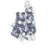
 Movie
Movie Controller


+ Open data

- Basic information
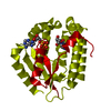
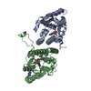
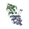
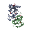
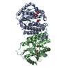
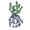
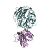
| Entry | Database: PDB / ID: 2no6 | ||||||
|---|---|---|---|---|---|---|---|
| Title | C4S dCK variant of dCK in complex with FTC+ADP | ||||||
 Components Components | deoxycytidine kinase | ||||||
 Keywords Keywords | TRANSFERASE / dCK / emtricitabine / human deoxycytidine kinase / FTC / enantiomer / antiviral | ||||||
| Function / homology |  Function and homology informationdeoxycytidine kinase / 2'-deoxyadenosine kinase / deoxyguanosine kinase / dAMP salvage / deoxycytidine kinase activity / nucleoside phosphate biosynthetic process / deoxyguanosine kinase activity / deoxyadenosine kinase activity / Pyrimidine salvage / cytidine kinase activity ...deoxycytidine kinase / 2'-deoxyadenosine kinase / deoxyguanosine kinase / dAMP salvage / deoxycytidine kinase activity / nucleoside phosphate biosynthetic process / deoxyguanosine kinase activity / deoxyadenosine kinase activity / Pyrimidine salvage / cytidine kinase activity / pyrimidine nucleotide metabolic process / Purine salvage / phosphorylation / protein homodimerization activity / mitochondrion / nucleoplasm / ATP binding / cytosol / cytoplasm Function and homology informationdeoxycytidine kinase / 2'-deoxyadenosine kinase / deoxyguanosine kinase / dAMP salvage / deoxycytidine kinase activity / nucleoside phosphate biosynthetic process / deoxyguanosine kinase activity / deoxyadenosine kinase activity / Pyrimidine salvage / cytidine kinase activity ...deoxycytidine kinase / 2'-deoxyadenosine kinase / deoxyguanosine kinase / dAMP salvage / deoxycytidine kinase activity / nucleoside phosphate biosynthetic process / deoxyguanosine kinase activity / deoxyadenosine kinase activity / Pyrimidine salvage / cytidine kinase activity / pyrimidine nucleotide metabolic process / Purine salvage / phosphorylation / protein homodimerization activity / mitochondrion / nucleoplasm / ATP binding / cytosol / cytoplasmSimilarity search - Function | ||||||
| Biological species |  Homo sapiens (human) Homo sapiens (human) | ||||||
| Method | X-RAY DIFFRACTION / SYNCHROTRON / MOLECULAR REPLACEMENT / Resolution: 1.9 Å | ||||||
 Authors Authors | Sabini, E. / Hazra, S. / Konrad, M. / Burley, S.K. / Lavie, A. | ||||||
 Citation Citation | Journal: J.Med.Chem. / Year: 2007 Title: Nonenantioselectivity Property of Human Deoxycytidine Kinase Explained by Structures of the Enzyme in Complex with l- and d-Nucleosides. Authors: Sabini, E. / Hazra, S. / Konrad, M. / Lavie, A. | ||||||
| History |
|
- Structure visualization
| Structure viewer | Molecule: MolmilJmol/JSmol |
|---|
- Downloads & links
-Download
| PDBx/mmCIF format | 2no6.cif.gz | 116.7 KB | Display | PDBx/mmCIF format |
|---|---|---|---|---|
| PDB format | pdb2no6.ent.gz | 88.8 KB | Display | PDB format |
| PDBx/mmJSON format | 2no6.json.gz | Tree view | PDBx/mmJSON format | |
| Others |  Other downloads Other downloads |
-Validation report
| Arichive directory | https://data.pdbj.org/pub/pdb/validation_reports/no/2no6ftp://data.pdbj.org/pub/pdb/validation_reports/no/2no6 | HTTPS FTP |
|---|
-Related structure data
| Related structure data | 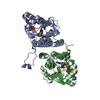 2no0C 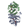 2no1C 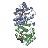 2no7C 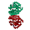 1p5zS S: Starting model for refinement C: citing same article ( |
|---|---|
| Similar structure data |


-Links
 PDBj
PDBj
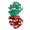
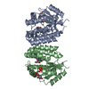
- Assembly
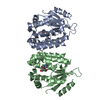
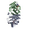
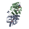
| Deposited unit | 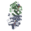
| ||||||||
|---|---|---|---|---|---|---|---|---|---|
| 1 |
| ||||||||
| 2 | 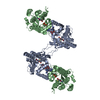
| ||||||||
| 3 | 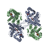
| ||||||||
| Unit cell |
| ||||||||
| Components on special symmetry positions |
|
-Components
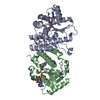
| #1: Protein | / dCK Mass: 32659.588 Da / Num. of mol.: 2 / Mutation: C9S,C45S,C59S,C146S Source method: isolated from a genetically manipulated source Source: (gene. exp.) Homo sapiens (human) / Gene: DCK / Plasmid: pET14b / Production host:  Escherichia coli (E. coli) / References: UniProt: P27707, deoxycytidine kinase Escherichia coli (E. coli) / References: UniProt: P27707, deoxycytidine kinase#2: Chemical | Adenosine diphosphate  Mass: 427.201 Da / Num. of mol.: 2 / Source method: obtained synthetically / Formula: C10H15N5O10P2 / Comment: ADP, energy-carrying molecule*YM Mass: 427.201 Da / Num. of mol.: 2 / Source method: obtained synthetically / Formula: C10H15N5O10P2 / Comment: ADP, energy-carrying molecule*YM#3: Chemical | Emtricitabine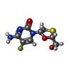  Mass: 247.247 Da / Num. of mol.: 2 / Source method: obtained synthetically / Formula: C8H10FN3O3S / Comment: medication, inhibitor*YM Mass: 247.247 Da / Num. of mol.: 2 / Source method: obtained synthetically / Formula: C8H10FN3O3S / Comment: medication, inhibitor*YM#4: Water | ChemComp-HOH / | Water Mass: 18.015 Da / Num. of mol.: 178 / Source method: isolated from a natural source / Formula: H2O Mass: 18.015 Da / Num. of mol.: 178 / Source method: isolated from a natural source / Formula: H2O |
|---|
-Experimental details
-Experiment
| Experiment | Method: X-RAY DIFFRACTION / Number of used crystals: 1 |
|---|
- Sample preparation
| Crystal | Density Matthews: 2.25 Å3/Da / Density % sol: 45.39 % |
|---|---|
| Crystal grow | Temperature: 298 K / Method: vapor diffusion, hanging drop / pH: 7.5 Details: Reservoir containing 0.95-1.5M trisodium citrate dihydrate and 100mM HEPES, pH 7.5., VAPOR DIFFUSION, HANGING DROP, temperature 298K |
-Data collection
| Diffraction | Mean temperature: 100 K |
|---|---|
| Diffraction source | Source: SYNCHROTRON / Site: APS  / Beamline: 22-BM / Wavelength: 1 Å / Beamline: 22-BM / Wavelength: 1 Å |
| Detector | Type: MARMOSAIC 225 mm CCD / Detector: CCD / Date: Nov 20, 2005 / Details: mirrors |
| Radiation | Monochromator: Rosenbaum-Rock monochromator high-resolution double-crystal Si (111) sagittal focusing, Rosenbaum-Rock vertical Protocol: SINGLE WAVELENGTH / Monochromatic (M) / Laue (L): M / Scattering type: x-ray |
| Radiation wavelength | Wavelength: 1 Å / Relative weight: 1 |
| Reflection | Resolution: 1.9→30 Å / Num. all: 43903 / Num. obs: 43616 / % possible obs: 99.3 % / Observed criterion σ(I): -3 / Redundancy: 10.1 % / Biso Wilson estimate: 28.5 Å2 / Rsym value: 0.087 / Net I/σ(I): 16.7 |
| Reflection shell | Resolution: 1.9→2 Å / Redundancy: 9.5 % / Mean I/σ(I) obs: 3.8 / Num. unique all: 6730 / Rsym value: 0.569 / % possible all: 96.4 |
- Processing
| Software |
| ||||||||||||||||||||||||||||||||||||||||||||||||||||||||||||||||||||||||||||||||||||||||||
|---|---|---|---|---|---|---|---|---|---|---|---|---|---|---|---|---|---|---|---|---|---|---|---|---|---|---|---|---|---|---|---|---|---|---|---|---|---|---|---|---|---|---|---|---|---|---|---|---|---|---|---|---|---|---|---|---|---|---|---|---|---|---|---|---|---|---|---|---|---|---|---|---|---|---|---|---|---|---|---|---|---|---|---|---|---|---|---|---|---|---|---|
| Refinement | Method to determine structure: MOLECULAR REPLACEMENT Starting model: 1P5Z Resolution: 1.9→30 Å / Cor.coef. Fo:Fc: 0.961 / Cor.coef. Fo:Fc free: 0.94 / SU B: 3.783 / SU ML: 0.11 / Cross valid method: THROUGHOUT / σ(F): 0 / ESU R: 0.157 / ESU R Free: 0.15 / Stereochemistry target values: MAXIMUM LIKELIHOOD / Details: HYDROGENS HAVE BEEN ADDED IN THE RIDING POSITIONS
| ||||||||||||||||||||||||||||||||||||||||||||||||||||||||||||||||||||||||||||||||||||||||||
| Solvent computation | Ion probe radii: 0.8 Å / Shrinkage radii: 0.8 Å / VDW probe radii: 1.4 Å / Solvent model: MASK | ||||||||||||||||||||||||||||||||||||||||||||||||||||||||||||||||||||||||||||||||||||||||||
| Displacement parameters | Biso mean: 31.416 Å2
| ||||||||||||||||||||||||||||||||||||||||||||||||||||||||||||||||||||||||||||||||||||||||||
| Refinement step | Cycle: LAST / Resolution: 1.9→30 Å
| ||||||||||||||||||||||||||||||||||||||||||||||||||||||||||||||||||||||||||||||||||||||||||
| Refine LS restraints |
| ||||||||||||||||||||||||||||||||||||||||||||||||||||||||||||||||||||||||||||||||||||||||||
| LS refinement shell | Resolution: 1.9→1.951 Å / Total num. of bins used: 20
|
