Myosinheavychain, non-muscleIIa / Myosin-9 / Cellular myosin heavy chain / type A / Myosin heavy chain 9 / Myosin heavy chain / non- ...Myosin-9 / Cellular myosin heavy chain / type A / Myosin heavy chain 9 / Myosin heavy chain / non-muscle IIa / Non-muscle myosin heavy chain A / NMMHC-A / Non-muscle myosin heavy chain IIa / NMMHC II-a / NMMHC-IIA
内容: 0.5 mM protein_1, 1 mM [U-100% 13C; U-100% 15N] protein_2, 5 mM CALCIUM ION, 20 mM MES, 20 mM sodium chloride, 4 mM TCEP, 95% H2O/5% D2O 溶媒系: 95% H2O/5% D2O
試料
濃度 (mg/ml)
構成要素
Isotopic labeling
Solution-ID
0.5mM
entity_1-1
1
1mM
entity_2-2
[U-100% 13C; U-100% 15N]
1
5mM
CALCIUM ION-3
1
20mM
MES-4
1
20mM
sodium chloride-5
1
4mM
TCEP-6
1
試料状態
イオン強度: 0.02 / pH: 6.1 / 圧: ambient / 温度: 313 K
-
NMR測定
NMRスペクトロメーター
タイプ
製造業者
モデル
磁場強度 (MHz)
Spectrometer-ID
Bruker Avance
Bruker
AVANCE
600
1
Bruker Avance
Bruker
AVANCE
800
2
-
解析
NMR software
名称
バージョン
開発者
分類
CCPN_Analysis
2.1
CCPN
chemicalshiftassignment
TopSpin
2.1
BrukerBiospin
解析
CNS
1.1
Brunger, Adams, Clore, Gros, NilgesandRead
構造決定
CNS
1.1
Brunger, Adams, Clore, Gros, NilgesandRead
精密化
ARIA
1.2
Linge, O'DonoghueandNilges
構造決定
ProcheckNMR
LaskowskiandMacArthur
geometryoptimization
精密化
手法: torsion angle dynamics / ソフトェア番号: 1
NMR constraints
NOE constraints total: 7085 / NOE intraresidue total count: 2709 / NOE long range total count: 2753 / NOE medium range total count: 1600 / NOE sequential total count: 1820 / Protein chi angle constraints total count: 0 / Protein other angle constraints total count: 0 / Protein phi angle constraints total count: 182 / Protein psi angle constraints total count: 182
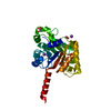
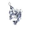
代表構造
選択基準: lowest energy
NMRアンサンブル
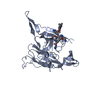
Average torsion angle constraint violation: 1.9 ° コンフォーマー選択の基準: structures with the lowest energy 計算したコンフォーマーの数: 200 / 登録したコンフォーマーの数: 20 / Maximum lower distance constraint violation: 0 Å / Maximum torsion angle constraint violation: 8 ° / Maximum upper distance constraint violation: 0.5 Å / Torsion angle constraint violation method: cns
NMR ensemble rms
Distance rms dev: 0.05 Å / Distance rms dev error: 0.004 Å
 ムービー
ムービー コントローラー


 データを開く
 基本情報
基本情報 要素
要素 キーワード
キーワード 機能・相同性情報
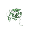
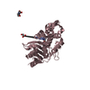
 Homo sapiens (ヒト)
 データ登録者
 引用
引用 構造の表示
構造の表示 ダウンロードとリンク
 その他のダウンロード
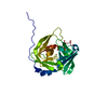
 PDBj
PDBj


 集合体
集合体

 試料調製
試料調製 解析
解析