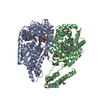
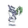
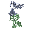
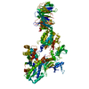
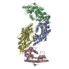
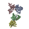
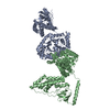
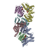
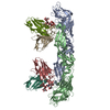
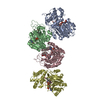
ジャーナル: J Am Chem Soc / 年: 2010 タイトル: Solution structure of the 128 kDa enzyme I dimer from Escherichia coli and its 146 kDa complex with HPr using residual dipolar couplings and small- and wide-angle X-ray scattering. 著者: Charles D Schwieters / Jeong-Yong Suh / Alexander Grishaev / Rodolfo Ghirlando / Yuki Takayama / G Marius Clore / 要旨: The solution structures of free Enzyme I (EI, ∼128 kDa, 575 × 2 residues), the first enzyme in the bacterial phosphotransferase system, and its complex with HPr (∼146 kDa) have been solved using ...The solution structures of free Enzyme I (EI, ∼128 kDa, 575 × 2 residues), the first enzyme in the bacterial phosphotransferase system, and its complex with HPr (∼146 kDa) have been solved using novel methodology that makes use of prior structural knowledge (namely, the structures of the dimeric EIC domain and the isolated EIN domain both free and complexed to HPr), combined with residual dipolar coupling (RDC), small- (SAXS) and wide- (WAXS) angle X-ray scattering and small-angle neutron scattering (SANS) data. The calculational strategy employs conjoined rigid body/torsion/Cartesian simulated annealing, and incorporates improvements in calculating and refining against SAXS/WAXS data that take into account complex molecular shapes in the description of the solvent layer resulting in a better representation of the SAXS/WAXS data. The RDC data orient the symmetrically related EIN domains relative to the C(2) symmetry axis of the EIC dimer, while translational, shape, and size information is provided by SAXS/WAXS. The resulting structures are independently validated by SANS. Comparison of the structures of the free EI and the EI-HPr complex with that of the crystal structure of a trapped phosphorylated EI intermediate reveals large (∼70-90°) hinge body rotations of the two subdomains comprising the EIN domain, as well as of the EIN domain relative to the dimeric EIC domain. These large-scale interdomain motions shed light on the structural transitions that accompany the catalytic cycle of EI.
履歴
登録
2010年4月29日
登録サイト: BMRB / 処理サイト: RCSB
改定 1.0
2010年9月15日
Provider: repository / タイプ: Initial release
改定 1.1
2011年7月13日
Group: Version format compliance
改定 1.2
2019年5月8日
Group: Data collection / Database references / カテゴリ: pdbx_database_related
内容: 20 mM TRIS, 100 mM sodium chloride, 10 mM DTT, 4 mM MgCl2, 1 mM EDTA, 10 % D2O, 1 tablet protease inhibitor, 0.15 mM EI dimer, 90% H2O/10% D2O 溶媒系: 90% H2O/10% D2O
試料
濃度 (mg/ml)
構成要素
Solution-ID
20mM
TRIS-1
1
100mM
sodium chloride-2
1
10mM
DTT-3
1
4mM
MgCl2-4
1
1mM
EDTA-5
1
10 %
D2O-6
1
1 %
protease inhibitor-7
1
0.15mM
EI dimer-8
1
試料状態
pH: 7.4 / 圧: ambient / 温度: 310 K
-
データ収集
NMRスペクトロメーター
タイプ: Bruker DRX / 製造業者: Bruker / モデル: DRX / 磁場強度: 800 MHz
Soln scatter
Data analysis software list: GNOM / Protein length: 150 / Sample pH: 7.4 / 温度: 298 K
タイプ
ID
Conc. range (mg/ml)
検出器タイプ
Mean guiner radius (nm)
Num. of time frames
Source beamline
Source class
Source type
x-ray
1
2.5-5
GoldCCD
41.9
20
2.12-IDC
Y
ALS
neutron
2
5
44.2
30MNG3
N
NISTNCNR
-
解析
NMR software
名称
バージョン
開発者
分類
Xplor-NIH
2.25
Schwieters, Kuszewski, TjandraandClore
構造決定
Xplor-NIH
2.25
Schwieters, Kuszewski, TjandraandClore
精密化
精密化
手法: simulated annealing / ソフトェア番号: 1 詳細: STRUCTURE STATISTICS: MODEL 1: SAXS CHI2 Q->0.44: 0.43 SAXS CHI2 FULL RANGE: 2.26 SANS CHI2: 0.39 RDC R-FACTOR: 18.03 % RDC DA: 14.5 HZ RDC RH: 0.49 MODEL 2: SAXS CHI2 Q->0.44: 0.40 SAXS CHI2 ...詳細: STRUCTURE STATISTICS: MODEL 1: SAXS CHI2 Q->0.44: 0.43 SAXS CHI2 FULL RANGE: 2.26 SANS CHI2: 0.39 RDC R-FACTOR: 18.03 % RDC DA: 14.5 HZ RDC RH: 0.49 MODEL 2: SAXS CHI2 Q->0.44: 0.40 SAXS CHI2 FULL RANGE: 1.99 SANS CHI2: 0.58 RDC R-FACTOR: 18.07 RDC DA: 14.5 HZ RDC RH: 0.48 AVERAGE OVER THE FULL 99-MEMBER ENSEMBLE: SAXS CHI2 Q->0.44: 0.29 +/- 0.05 SAXS CHI2 FULL RANGE: 1.34 +/- 0.27 SANS CHI2: 0.41 +/- 0.08 RDC R-FACTOR: 18.07 +/- 0.02 % RDC DA: 14.5 +/- 0.1 HZ RDC RH: 0.49 +/- 0.00
代表構造
選択基準: closest to the average
NMRアンサンブル
コンフォーマー選択の基準: target function / 計算したコンフォーマーの数: 120 / 登録したコンフォーマーの数: 2
Soln scatter model
コンフォーマー選択の基準: REGULARIZED MEAN OF 99 MODELS 詳細: The initial structure of the EI dimer was constructed as a hybrid of the crystal structure of phosphorylated EI intermediate captured by the inhibitor oxalate (PDB code 2HWG) and the NMR ...詳細: The initial structure of the EI dimer was constructed as a hybrid of the crystal structure of phosphorylated EI intermediate captured by the inhibitor oxalate (PDB code 2HWG) and the NMR structure of the EIN-HPr complex (PDB code 3EZA). Throughout the structure determination, the backbone atomic coordinates of each EIN domain (residues 1-254) were treated as rigid bodies, with the two symmetry related EIC domains (residues 262- 573) held fixed in space. Coordinates in the linker region (residues 255-261) were allowed varying degrees of freedom during the calculation through the use of the internal variable module (IVM) of Xplor-NIH. This entry corresponds to the regularized mean of the 99 structures for which data was reported in the primary publication, with the B-factor column representing the per-atom spread (in B-factor units). The calculated structural statistics for the original 99 structures and for the regularized mean are shown below. Entry fitting list: PDB ENTRIES 2HWG AND 3EZA / Num. of conformers calculated: 99 / Num. of conformers submitted: 1 / 代表コンフォーマー: 1 / Software list: GNOM,XPLOR-NIH
 ムービー
ムービー コントローラー


 データを開く
 基本情報
基本情報 要素
要素 Phosphoenolpyruvate—protein phosphotransferase
  キーワード
キーワード 機能・相同性情報
 データ登録者
 引用
引用
 構造の表示
構造の表示 ダウンロードとリンク
 その他のダウンロード
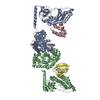


 PDBj
PDBj
 集合体
集合体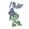
 試料調製
試料調製 解析
解析