 Movie
Movie Controller


+ Open data

- Basic information
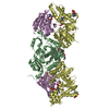
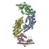
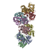
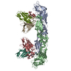
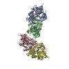
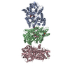
| Entry | Database: PDB / ID: 6s84 | |||||||||
|---|---|---|---|---|---|---|---|---|---|---|
| Title | TsaBDE complex from Thermotoga maritima | |||||||||
 Components Components |
| |||||||||
 Keywords Keywords |  RNA BINDING PROTEIN / modification / t6A / tRNA maturation RNA BINDING PROTEIN / modification / t6A / tRNA maturation | |||||||||
| Function / homology |  Function and homology information Function and homology informationN6-L-threonylcarbamoyladenine synthase / N(6)-L-threonylcarbamoyladenine synthase activity / tRNA threonylcarbamoyladenosine modification / iron ion binding / cytosol / cytoplasmSimilarity search - Function | |||||||||
| Biological species |   Thermotoga maritima (bacteria) Thermotoga maritima (bacteria) | |||||||||
| Method | X-RAY DIFFRACTION / SYNCHROTRON / MOLECULAR REPLACEMENT / Resolution: 2.89 Å | |||||||||
 Authors Authors | Missoury, S. / Li-de-La-Sierra-Gallay, I. / van Tilbeurgh, H. | |||||||||
 Citation Citation | Journal: Nucleic Acids Res. / Year: 2018 Title: The structure of the TsaB/TsaD/TsaE complex reveals an unexpected mechanism for the bacterial t6A tRNA-modification. Authors: Missoury, S. / Plancqueel, S. / Li de la Sierra-Gallay, I. / Zhang, W. / Liger, D. / Durand, D. / Dammak, R. / Collinet, B. / van Tilbeurgh, H. | |||||||||
| History |
|
- Structure visualization
| Structure viewer | Molecule: MolmilJmol/JSmol |
|---|
- Downloads & links
-Download
| PDBx/mmCIF format | 6s84.cif.gz | 278.6 KB | Display | PDBx/mmCIF format |
|---|---|---|---|---|
| PDB format | pdb6s84.ent.gz | 220.9 KB | Display | PDB format |
| PDBx/mmJSON format | 6s84.json.gz | Tree view | PDBx/mmJSON format | |
| Others |  Other downloads Other downloads |
-Validation report
| Arichive directory | https://data.pdbj.org/pub/pdb/validation_reports/s8/6s84ftp://data.pdbj.org/pub/pdb/validation_reports/s8/6s84 | HTTPS FTP |
|---|
-Related structure data
| Similar structure data |
|---|


-Links
 PDBj
PDBj


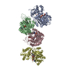
- Assembly
| Deposited unit | 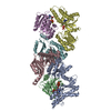
| ||||||||
|---|---|---|---|---|---|---|---|---|---|
| 1 |
| ||||||||
| Unit cell |
|
-Components
-Protein , 3 types, 6 molecules ADEBCF
| #1: Protein | Mass: 35672.203 Da / Num. of mol.: 2 Source method: isolated from a genetically manipulated source Source: (gene. exp.) Thermotoga maritima (strain ATCC 43589 / MSB8 / DSM 3109 / JCM 10099) (bacteria)Strain: ATCC 43589 / MSB8 / DSM 3109 / JCM 10099 / Gene: tsaD, gcp, TM_0145 / Production host: Escherichia coli (E. coli)References: UniProt: Q9WXZ2, N6-L-threonylcarbamoyladenine synthase #2: Protein | Mass: 21513.488 Da / Num. of mol.: 2 Source method: isolated from a genetically manipulated source Source: (gene. exp.) Thermotoga maritima (strain ATCC 43589 / MSB8 / DSM 3109 / JCM 10099) (bacteria)Strain: ATCC 43589 / MSB8 / DSM 3109 / JCM 10099 / Gene: Tmari_1641 / Production host: Escherichia coli (E. coli) / References: UniProt: R4NRX5#3: Protein | Mass: 23020.842 Da / Num. of mol.: 2 Source method: isolated from a genetically manipulated source Source: (gene. exp.) Thermotoga maritima (strain ATCC 43589 / MSB8 / DSM 3109 / JCM 10099) (bacteria)Strain: ATCC 43589 / MSB8 / DSM 3109 / JCM 10099 / Gene: tsaB, TM_0874, Tmari_0876 / Production host: Escherichia coli (E. coli) / References: UniProt: Q9WZX7 |
|---|
-Non-polymers , 4 types, 25 molecules 


| #4: Chemical | ChemComp-APC /  Mass: 505.208 Da / Num. of mol.: 4 / Source method: isolated from a natural source / Formula: C11H18N5O12P3 / Comment: AMP-CPP, energy-carrying molecule analogue*YM Mass: 505.208 Da / Num. of mol.: 4 / Source method: isolated from a natural source / Formula: C11H18N5O12P3 / Comment: AMP-CPP, energy-carrying molecule analogue*YM#5: Chemical |  Mass: 24.305 Da / Num. of mol.: 2 / Source method: obtained synthetically / Formula: Mg Mass: 24.305 Da / Num. of mol.: 2 / Source method: obtained synthetically / Formula: Mg#6: Chemical | ChemComp-LEU / | Leucine Type: L-peptide linking / Mass: 131.173 Da / Num. of mol.: 1 / Source method: obtained synthetically / Formula: C6H13NO2 Type: L-peptide linking / Mass: 131.173 Da / Num. of mol.: 1 / Source method: obtained synthetically / Formula: C6H13NO2#7: Water | ChemComp-HOH / | WaterMass: 18.015 Da / Num. of mol.: 18 / Source method: isolated from a natural source / Formula: H2O |
|---|
-Experimental details
-Experiment
| Experiment | Method: X-RAY DIFFRACTION / Number of used crystals: 1 |
|---|
- Sample preparation
| Crystal | Density Matthews: 2.63 Å3/Da / Density % sol: 53.27 % |
|---|---|
| Crystal grow | Temperature: 293 K / Method: vapor diffusion, sitting drop / Details: 13 % PEG 8000, 0.1 mM Imidazole pH 7 |
-Data collection
| Diffraction | Mean temperature: 100 K / Serial crystal experiment: N | ||||||||||||||||||||||||||||||||||||||||||||||||||||||||||||||||||||||||||||||||||||||||||||||||||||
|---|---|---|---|---|---|---|---|---|---|---|---|---|---|---|---|---|---|---|---|---|---|---|---|---|---|---|---|---|---|---|---|---|---|---|---|---|---|---|---|---|---|---|---|---|---|---|---|---|---|---|---|---|---|---|---|---|---|---|---|---|---|---|---|---|---|---|---|---|---|---|---|---|---|---|---|---|---|---|---|---|---|---|---|---|---|---|---|---|---|---|---|---|---|---|---|---|---|---|---|---|---|
| Diffraction source | Source: SYNCHROTRON / Site: SOLEIL  / Beamline: PROXIMA 1 / Wavelength: 0.97857 Å / Beamline: PROXIMA 1 / Wavelength: 0.97857 Å | ||||||||||||||||||||||||||||||||||||||||||||||||||||||||||||||||||||||||||||||||||||||||||||||||||||
| Detector | Type: PSI PILATUS 6M / Detector: PIXEL / Date: Aug 7, 2018 | ||||||||||||||||||||||||||||||||||||||||||||||||||||||||||||||||||||||||||||||||||||||||||||||||||||
| Radiation | Protocol: SINGLE WAVELENGTH / Monochromatic (M) / Laue (L): M / Scattering type: x-ray | ||||||||||||||||||||||||||||||||||||||||||||||||||||||||||||||||||||||||||||||||||||||||||||||||||||
| Radiation wavelength | Wavelength: 0.97857 Å / Relative weight: 1 | ||||||||||||||||||||||||||||||||||||||||||||||||||||||||||||||||||||||||||||||||||||||||||||||||||||
| Reflection | Resolution: 2.89→48.43 Å / Num. obs: 36847 / % possible obs: 99.1 % / Redundancy: 8.793 % / Biso Wilson estimate: 80.486 Å2 / CC1/2: 0.997 / Rmerge(I) obs: 0.187 / Rrim(I) all: 0.199 / Χ2: 1.18 / Net I/σ(I): 9.17 / Num. measured all: 323984 | ||||||||||||||||||||||||||||||||||||||||||||||||||||||||||||||||||||||||||||||||||||||||||||||||||||
| Reflection shell | Diffraction-ID: 1
|
- Processing
| Software |
| |||||||||||||||||||||||||||||||||||||||||||||
|---|---|---|---|---|---|---|---|---|---|---|---|---|---|---|---|---|---|---|---|---|---|---|---|---|---|---|---|---|---|---|---|---|---|---|---|---|---|---|---|---|---|---|---|---|---|---|
| Refinement | Method to determine structure: MOLECULAR REPLACEMENT / Resolution: 2.89→48.43 Å / Cor.coef. Fo:Fc: 0.954 / Cor.coef. Fo:Fc free: 0.904 / SU B: 28.035 / SU ML: 0.465 / Cross valid method: THROUGHOUT / σ(F): 0 / ESU R Free: 0.46 / Details: U VALUES : REFINED INDIVIDUALLY
| |||||||||||||||||||||||||||||||||||||||||||||
| Solvent computation | Ion probe radii: 0.8 Å / Shrinkage radii: 0.8 Å / VDW probe radii: 1.2 Å | |||||||||||||||||||||||||||||||||||||||||||||
| Displacement parameters | Biso max: 227.34 Å2 / Biso mean: 89.656 Å2 / Biso min: 32.6 Å2
| |||||||||||||||||||||||||||||||||||||||||||||
| Refinement step | Cycle: final / Resolution: 2.89→48.43 Å
| |||||||||||||||||||||||||||||||||||||||||||||
| Refine LS restraints |
| |||||||||||||||||||||||||||||||||||||||||||||
| LS refinement shell | Resolution: 2.894→2.969 Å / Rfactor Rfree error: 0 / Total num. of bins used: 20
|
